
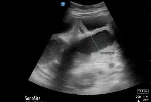
The Image of the Week comes to us from Drs. Kahra Nix, Ian Dodson, Darin Williams, and Jeremy Ackermann who used ultrasound to evaluate a 50 yo patient with COPD who presented with ROSC following PEA arrest. Can you identify the finding below? Thanks for all your great images this week. Happy Scanning! =============================== …
Category: Image of the Week
Jun 01
Image of the Week – Traumatic Arrest After GSW
The Image of the Week comes to us from Drs. Almebash and Reynolds who performed a FAST exam in a patient who presented in traumatic arrest following a GSW. Can you identify the pathology present? Thanks for all of your great images this week! Happy Scanning! =============================== Sierra Beck MD Assistant Professor Department of …
Apr 30
Image of the Week – Achilles Tendon Rupture
The Image of the Week comes to us from Drs Kahra Nix, Jose Rosa, and Jeremy Whitley & MS4s Eli and Rachel who used ultrasound to evaluate an athletic young man who experienced pain and a pop in his ankle whlle playing basketball. Can you identify the normal and abnormal structures below? Thanks for …
Apr 21
Image of the Week – Aortic Dissection
The Image of the Week comes to us from Drs. Kahra Nix, David Zhou, and Shikha Kapil who used ultrasound to evaluate a patient who presented after clinic visit for cough and an abnormal CXR prompted further work up. Can you interpret the image below that was obtained when the patient arrived in the ED? …
Apr 15
Image of the Week – Hepatic Abscess
The Image of the Week comes to us from Dr Layne Madden who used ultrasound to evaluate a patient who presented with fatigue, anorexia, and fever associated with epigastric pain on exam. Can you interpret the image below? Thanks for all of your great images this week! Happy Scanning! =============================== Sierra Beck MD Assistant …
Apr 01
Image of the Week – Superficial and Deep Peroneal Nerve Block
The Image of the Week comes to us from Drs. Karen Bowers and Mene Demestihas who used ultrasound to assist with anesthesia to a complex laceration involving the dorsum of the foot and first web space. They performed two nerve blocks, one of which is shown below. How would you anesthetize a wound like this? …
Mar 25
Image of the Week – Intussusception
The image of the week this week comes to us from Drs. Brandi Gunn and Jeff Siegelman who used bedside ultrasound to evaluate an adult with nausea and abdominal pain. They obtained and correctly interpreted the image below. Can you make the diagnosis? Thanks for all of your great images this week! Happy Scanning! =============================== …
Mar 21
Image of the Week – Appendicitis
This week’s Image of the Week features images from two patients. The first seen by Dr’s Meloy and Sizemore, then second by Dr’s Shah and Middlebrooks. Both presented with right lower quadrant pain. Take a look at the images and see if you can identify the pathology. Both of these patients were diagnosed with appendicitis. …
Mar 12
Image of the Week – Small Bowel Obstruction
Courtesy of Dr Wetendorf: This week’s image is brought to us by medical students Robbins and Lee. The patient presented with hypotension and emesis. Bedside ultrasound quickly captured the image below. Video captures several signs of small bowel obstruction (SBO):the classic “keyboard” sign (visualization of the plicae circulares), abnormal (bidirectional)peristalsis, and dilated loops of bowel. To …
Feb 29
Image of the Week – Liver GSW
Image of the Week courtesy of Dr Sierra Beck: The image of the week comes to us from Drs Pearl Ann Arnovitz and Todd Taylor who used ultrasound to evaluate a patient with a GSW to the right thoracoabdomen. Can you identify the pathology present? Thanks for all of your great images this week! Happy …
- 1
- 2