Latest News


Salaita lab in the news

Check out these videos to learn more about what we do!


NIH funds Emory center to advance cellular mechanics

“We are catalyzing the process of spreading our technology so that studying biomechanics becomes common and routine in biology”

Merck Prize boosts work on air sensor for pandemic pathogens

Chemist Khalid Salaita leads Emory students and faculty in a visionary project

'Firefly' imaging method zooms in on 'the forces within us'

Emory chemists make cellular forces visible at the molecular scale

New tool to analyze blood platelets holds major medical potential

A novel technique to test platelet function within a person’s blood sample is faster, easier and more precise than methods currently in use, an experimental study shows

Chameleon-inspired 'smart skin' materials that change color on demand

Hydrogels that dissipate mechanical strain do the trick!

Our Lab invented molecular tension fluorescence microscopy

The first approach to map cell traction forces with pN sensitivity! 

tension.jpeg

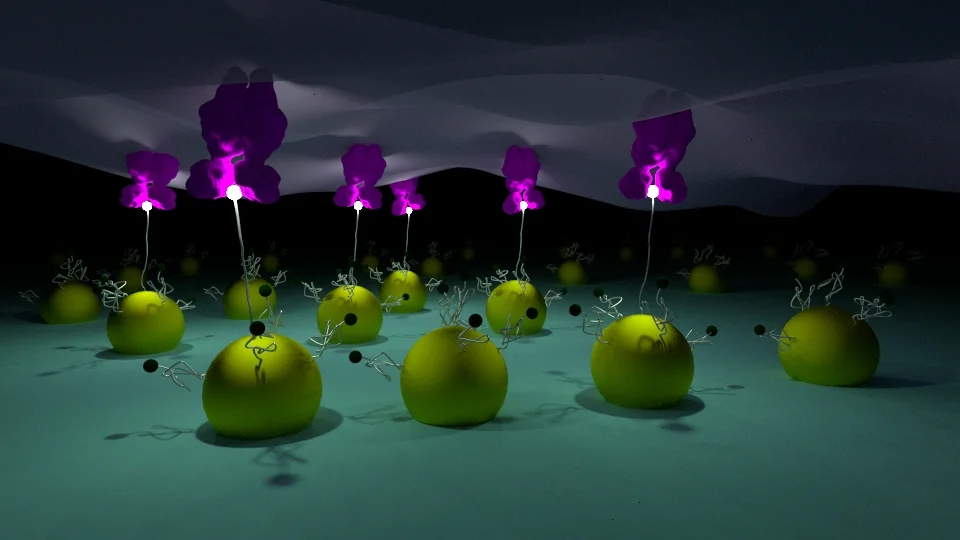
Our lab developed molecular tension fluorescence microscopy which has the potential to transform the study of cell biology by developing the tools to visualize molecular forces with the speed, convenience, and precision of conventional fluorescence microscopy. There is intense interest in studying cellular mechanobiology for a broad range of motivations that span from fundamental developmental biology to cancer diagnostics. For example, stem cells have been shown to feel and respond to the stiffness of the underlying substrate by steering differentiation based on the mechanical properties of the cellular microenvironment. 

We first reported these tension probes in Nature Methods in 2011. The design is fairly simple, and is akin to a macroscopic force gauge. Probes consist of a flexible linker (DNA, protein, polymer) flanked by a fluorophore and quencher. Probes are immobilized onto a substrate and present a biological ligand that engages the receptor of interest. When a cell applies a specific pN forces to stretch the probe, the fluorophore is separated from the quencher, thus leading to an enhancement in signal (up to 100 fold enhancement). 

INTEGRIN-MEDIATED MECHANOTRANSDUCTION

Integrins are receptors that span the plasma membrane and anchor cells to the external environment. We are interested in studying the interplay between mechanical forces and chemical signaling within integrins-mediated adhesions. Our recent data infers that a subset of integrin receptors applies >100 pN tensions, which is many fold greater than that of previously reported in literature, within focal adhesions of rat embryonic fibroblasts.


T-Cell Receptor Mechanotransduction

T cells protect the body against pathogens and cancer by recognizing specific foreign peptides on the cell surface. Because antigen recognition occurs at the junction between a migrating T cell and an antigen-presenting cell (APC), it is likely that cellular forces are generated and transmitted through T-cell receptor (TCR)-ligand bonds. In this project, we are employing DNA-based nanoparticle tension sensors producing the first molecular maps of TCR-ligand forces during T cell activation. We find that TCR forces are orchestrated in space and time, requiring the participation of CD8 co-receptor and adhesion molecules. Loss or damping of TCR forces results in weakened antigen discrimination, showing that T cells harness mechanics to optimize the specificity of response to ligand.

Mechanically Induced Catalytic Amplification Reaction for Readout of Cellular Forces

Mechanics play a fundamental role in cell biology, but detecting piconewton (pN) forces is challenging because of a lack of accessible and high throughput assays. In this project, we are developing mechanically induced catalytic amplification reactions (MCR) for readout of cell forces. As a proof of concept, the assay was used to test the activity of a mechanomodulatory drugs and integrin adhesion receptor antibodies. To the best of our knowledge, this is the first example of a catalytic reaction triggered in response to molecular piconewton forces. The MCR may transform the field of mechanobiology by providing a new facile tool to detect receptor specific mechanics with the convenience of the polymerase chain reaction (PCR).


Optomechanical actuator: New Tool to manipulate mechanics at the nanoscale using light

The research in this subgroup focuses on the development of active materials that can be triggered to deliver specific pN forces. These forces can then be used to drive biochemical reactions in living cells or alternatively to trigger a chemical transformation in soft materials. The center-piece of this effort focuses on the optomechanical actuator nanoparticle (see TEM  to the left). These particles are comprised of a plasmonic gold nanorod coated with a responsive polymer film. Upon illumination of the particles, the gold rod becomes hot, which drives the collapse of the polymer shell.  


Picture1.png
untitled2.png

Manipulating Mechanics in living cells

The majority of cells within multicellular organisms experience forces that are highly orchestrated in space and time. Several methods make it possible to investigate the cellular response to spatially confined mechanical inputs. For example, micropipettes and single-molecule techniques are used to physically prod the apical side of cells, but such approaches have low throughput. Magnetic actuation of nanoparticles and micropillars can trigger mechanotransduction pathways but require either a sparse density of magnetic elements or sophisticated microfabricated structures that focus an external magnetic field. Therefore, magnetic stimulation of mechanotransduction circuits remains specialized and is not widely used. Manipulating forces with molecular specificity and high spatiotemporal resolution remains a hurdle.

Optical approaches for the manipulation of biological systems are rapidly proliferating, as exemplified by caged or photoswitchable molecules and by optogenetic constructs. Similarly, methods to harness light for delivering precise physical inputs to biological systems could potentially transform the study of mechanotransduction.

Toward this goal, we used our optomechanical actuator nanoparticles to manipulate receptor mechanics with high spatiotemporal resolution using near-infrared (NIR) illumination. Nanoparticles shrink rapidly upon illumination, thereby applying a mechanical load to receptor-ligand complexes decorating the immobilized particle. The NIR optical pulse train controls the amplitude, duration, repetition and loading rate of mechanical input. Nanoparticles are immobilized onto standard glass coverslips, allowing cell imaging and manipulation using a conventional optical microscope equipped with an inexpensive NIR laser. Therefore, cell response to mechanical forces can be characterized with unprecedented spatial and temporal resolution. 

Picture1.png
 

Conditional DNA Therapeutics for Gene Regulation

Because DNA–nanoparticles (NPs) and antisense oligonucleotides (ASOs) are internalized by many cell types, they offer limited control in terms of tissue or cell type specificity. To take a step toward solving this issue, we incorporated toehold-mediated strand exchange, a versatile molecular programming modality, to switch the DNA-based therapeutics from an inactive state to an active state in the presence of a specific RNA input. Because many transcripts are unique to cell subtype or disease state, this approach could one day lead to responsive nucleic acid therapeutics with enhanced specificity.

Multivalent-Binding DNA Nanoparticles

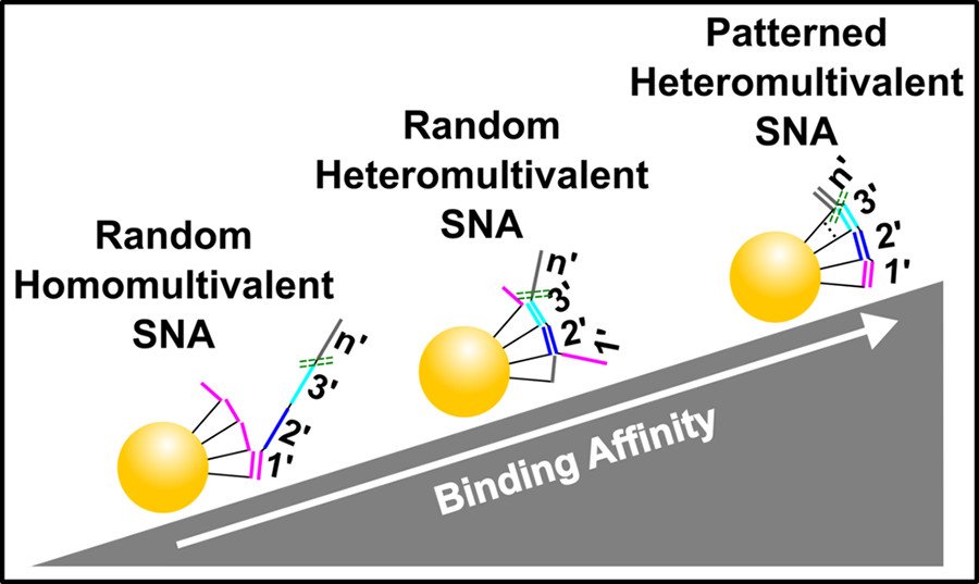
Improving the affinity of nucleic acids to their complements is an important goal in antisense therapy and diagnostics. One potential approach to achieving this goal is to use multivalent binding, which often boosts the affinity between ligands and receptors, as exemplified by virus–cell binding. We thus investigated the binding of heteromultivalent DNA–nanoparticle conjugates, and by developing a method to spatially pattern oligonucleotides on a particle, we demonstrated that the molecular organization is critical for effective binding.

LIPID-Nanostructures as scaffolds for DNA Delivery

Delivery of nucleic acids can be hindered by nuclease susceptibility, endosome trapping, and clearance. Multiple nanotechnology scaffolds have offered promising solutions, and among these, lipid-based systems are advantageous because of their high biocompatibility and low toxicity. However, many lipid nanoparticle systems still have issues regarding stability, rapid clearance, and cargo leakage. We thus demonstrated the use of a synthetic nanodisc (ND) scaffold functionalized with an antisense oligonucleotide (ASO) to reduce mRNA transcript levels, potentially offering an improved platform for ASO therapeutics.

 
roller.jpeg
 

Biological nanomotors are ubiquitous throughout many vital processes such as meiosis, mitosis, and cellular transport. They can walk >10 um at rates of 10 um/s; yet, at best, synthetic nanomachines can only walk 100 nm with a speed of 0.1 nm/s. Our goal is to improve the capabilities of synthetic motors so we can design more sophisticated materials. We developed micron and nano-sized DNA-based machines that roll rather than walk, and consequently have a maximum speed and processivity that is three orders of magnitude greater than the maximum for conventional DNA motors. The motors are made from DNA-coated spherical particles or rod-shaped DNA origami that hybridize to a surface modified with complementary RNA. The motion is achieved through the addition of RNase H, which selectively hydrolyses the hybridized RNA. The motors can move in a self-avoiding manner, and anisotropic particles, such as dimerized or rod-shaped particles, can travel linearly without a track or external force. These DNA-based machines generate 100+pN of force by converting chemical energy into controlled motion and could be of use in applications such as next-generation sensors, drug-delivery platforms and biological computing.

Screen Shot 2017-03-19 at 7.03.57 PM.png

Khalid Salaita

Samuel Candler Dobbs Professor
Emory University Chemistry Department

Khalid obtained his Ph.D. in the research group of Prof. Chad A. Mirkin at Northwestern University in 2006. During that time, he studied the electrochemical properties of organic adsorbates patterned onto gold films and developed massively parallel scanning probe lithography approaches. Khalid then started his postdoctoral training with Prof. Jay T. Groves at the University of California, Berkeley. He joined the faculty of Emory University as an assistant professor in the Department of Chemistry in 2009 and was promoted to associate professor in 2015. Khalid started his own lab at Emory University, where he currently investigates biophysical aspects of receptor-mediated cell signaling. Khalid is also a program faculty in the Department of Biomedical Engineering at Georgia Tech and Emory, and a program member of Cancer Cell Biology at Winship Cancer Institute. In recognition of his independent work, Khalid has received a number of awards, most notably: the Alfred Sloan Research Fellowship, the Camille-Dreyfus Teacher Scholar award, the NSF Early award, and the Kavli Fellowship. Khalid’s program is supported by NSF, NIH, and DARPA

See Khalid's CV and video interview for more details.