
Last week, the class attempted to visit the Musee des Moulages, but we were turned away as a result of a scheduling error. Our tickets were for the “7th” of June rather than the “1st.” Today, we bravely made the trek back to see the unique collection of wax dermatological models.
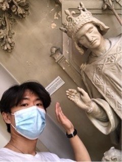
The Hospital Saint-Louis was ordered to be built by Henri IV who wanted plague victims to be treated outside the walls of the capital. I was able to get a selfie with him 🙂
This same hospital eventually hosted the genius of Jean-Louis Alibert who introduced the new medical discipline of dermatology. The idea to represent pathologies in three-dimensional representations was inspired by Jules Baretta who made models of fruit from paper board. According to the museum guide, Baretta made his first wax dermatological model in 1867, and eventually, the museum of wax models was established as a learning tool for the students of the hospitals.
Apparently, the Hospital Saint-Louis models were the inspiration for similar museums across the world and established a reputation that manifested in over 4,800 pieces today–the largest collection of its kind in the world. It is interesting to think that the majority of visualizations that students have access to today are primarily digital, and it may even be the case that the future will allow augmented reality renditions of these models.
Walking around the exhibit, there were many genital representations marred by a variety of disfigurations. I had a difficult time discerning what the diseases were (the descriptions were in French and there was no tour or explanation), but I definitely saw multiple sections dedicated to syphilis. This closely relates to our class discussion of syphilis wherein the disease presents itself in the skin as well as the forms of neuro and optical forms distinguished in our paper.
“The first symptom of primary syphilis is a usually painless open sore called a chancre (pronounced “shanker”). The chancre can appear within 10 days to 3 months (usually 2 to 6 weeks) after exposure.” As such, there were plenty of wax models of chancres throughout the exhibit, but I was not allowed to take any pictures as the majority of them were on the first floor by the supervisors.
However, on the second floor, I had a little more liberty to take a picture for the benefit of the class–a notable exhibit I saw was one dedicated to “abnormalities,” and I saw a model of a human “cyclops” baby. Again, the museum had a no photography policy, but I took one for the class, for science.
[Warning, graphic image below]
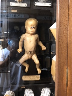
Here is what I found in research about cyclopia:
“Cyclopia (also cyclocephaly or synophthalmia) is a rare form of holoprosencephaly and is a congenital disorder (birth defect) characterized by the failure of the embryonic prosencephalon to properly divide the orbits of the eye into two cavities. It is the severest facial expression of the holoprosencephaly syndrome.1 Its incidence is 1 in 100 000 in newborns.”
This condition is interesting to read through especially considering our discussion of ‘ocular’ syphilis. There is little knowledge on the mechanisms or vision with cyclopia given the extremely low survival rates, and I was unable to find any case studies on adult cyclopia.
Overall, the visit was interesting in that I was introduced to a variety of ailments that I was not aware of, complete with visual representations.
References: