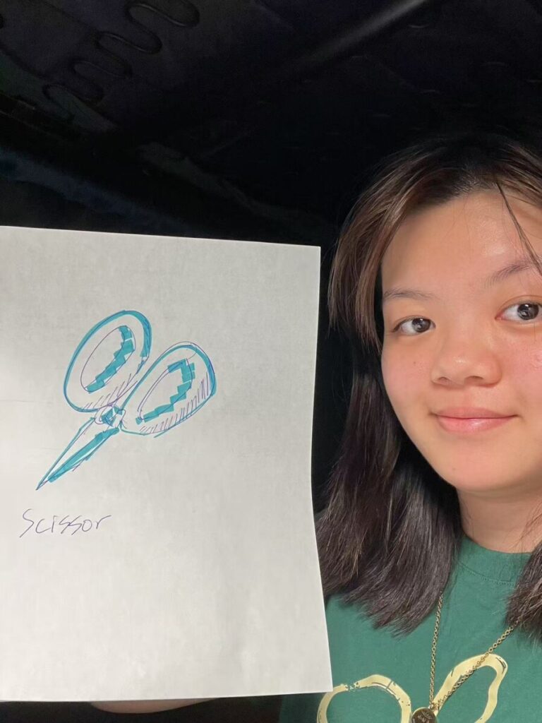
Last winter break, I was playing a game with my friends while waiting for dinner. One person draws a word using shapes and colors, and the other people guess what it is. We drew a lot of different objects, such as cars, cartoon characters, famous persons and a lot of different objects. The most fascinating thing is that, though the person who was drawing might not be the finest artist, we can still extract the important features from those things and being able to recognize them. For the below is a drawing of a scissor, and my friends can identify them very quickly.
Humans have a very profound ability to recognize things simply by looking at their components. For example, you are capable of recognizing it is a cat through a magnified picture by only seeing half of the head with the furs. Even if you have a different cat with different fur color and body shape, you are still able to recognize that, overall, this is a cat. This process is called object recognition, which we are able to identify the category of an object. While the light of such object falls into your retina with various shapes and colors, the key for recognition is to extract the matching representations that was stored in your memory. This is related to the perceptual memory, which involves brain regions such as the hippocampus, medial prefrontal cortex, parietal lobe, and occipital lobe (Chun and Johnson 2011 [1]).
While in the game, there are rounds that I have to recognize something I have fairly met before. I couldn’t make a connection of the art to the cue word which made me lose the game. While I was doubting if I had lost part of my memories for somehow because I indeed met the thing she drew before, she reminds me that a brain disorder we learned in Psychology class has similar behavior to my “symptom”. Such disorder is called Agnosia.
Agnosia is a syndrome that leads to failure of visual recognition. Which when you are presenting an object to the patient, the patient may not be able to relate it to the name of the object (Kumar and Wroten 2019 [4]). There is one case study done by José M. Ferro and Maria that infers the associative visual agnosia. This patient is a 58-year-old male with a temporo-occipital infarct, that includes the hippocampus, Para-hippocampal and fusiform gyri, calcarine cortex and temporo-occipital white matter. These regions are normally correlated with functions of storing long-term memory and integrating memories. The patient has normal ability of speech and aural comprehension if the order does not involve object manipulation. He has severe impaired reading ability and defect in recent verbal memory. The patient has defected ability to visually name the objects. As part of the study, he was asked to draw a particular object, though he could draw normally and instantaneously, the patient did not recognize the stimuli. The below is the drawing of this patient (Ferro and Santos 1984 [3]).

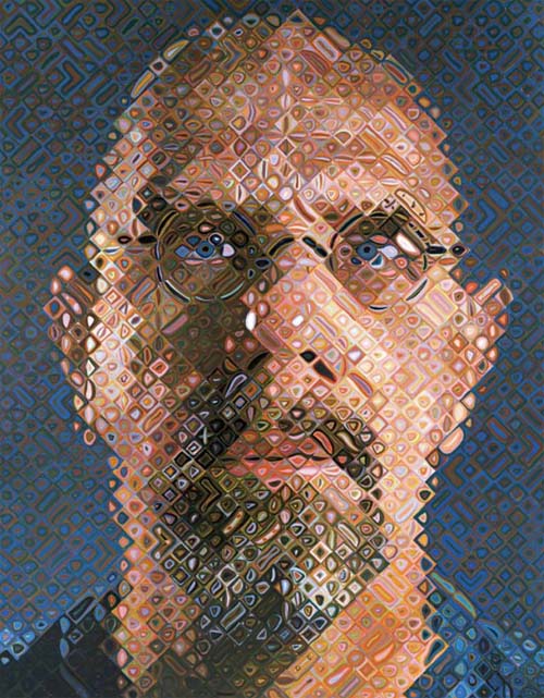
One subset of Agnosia is Prosopagnosia, which the person is incapable to recognize faces. Usually when we recognize faces, our temporal lobe and occipital face area plays a crucial role in face recognition. Chuck Close is a well-known artist diagnosed with prosopagnosia. However, his adversity flourishes his artistical expression. “Once I change the face into a two-dimensional object, I can commit it to memory. I have a photographic memory for things that are two-dimensional,” Close says. This conversion of face recognition using implicit memories switches to explicit memories helps him make facial recognition. This inspired his artwork. He expresses his sensation to human faces through abstract art that blends the photographs like looking through grids of glossed mirror (Farley 2011 [2]).
References:
[1] Chun, Marvin M., and Marcia K. Johnson. “Memory: Enduring Traces of Perceptual and Reflective Attention.” Neuron, vol. 72, no. 4, Nov. 2011, pp. 520–535, https://doi.org/10.1016/j.neuron.2011.10.026.
[2] Farley, Todd. “Disabilities Are at the Heart of Chuck Close’s Art.” Www.brainandlife.org, Sept. 2011, www.brainandlife.org/articles/dyslexia-paralysis-face-blindness-nothing-comes-between-legendary-artist-chuck.
[3] Ferro, José M., and Maria Emília Santos. “Associative Visual Agnosia: A Case Study.” Cortex, vol. 20, no. 1, Mar. 1984, pp. 121–134, https://doi.org/10.1016/s0010-9452(84)80029-5.
[4] Kumar, Anil, and Michael Wroten. “Agnosia.” National Library of Medicine, StatPearls Publishing, 11 Sept. 2019, www.ncbi.nlm.nih.gov/books/NBK493156/.