Noah Okada

Polyglot or Noob?

by Kaitlynn Love

art by Anonymous

Language is one of the primary ways we communicate with each other, and it is a fundamental aspect of human social behavior that is rapidly acquired early in life. Languages can be sound, sign, and tactile based, and language acquisition mechanisms differ depending on the time period during which the acquisition occurs. Second language acquisition may be more difficult for individuals due to the alternative mechanisms that are used beyond the sensitive period, a time span during which a young individual’s brain is especially malleable and shaped by experiences. Understanding the differences between the mechanisms used during versus beyond this period may be helpful in explaining some of the specific challenges involved with second language acquisition. 


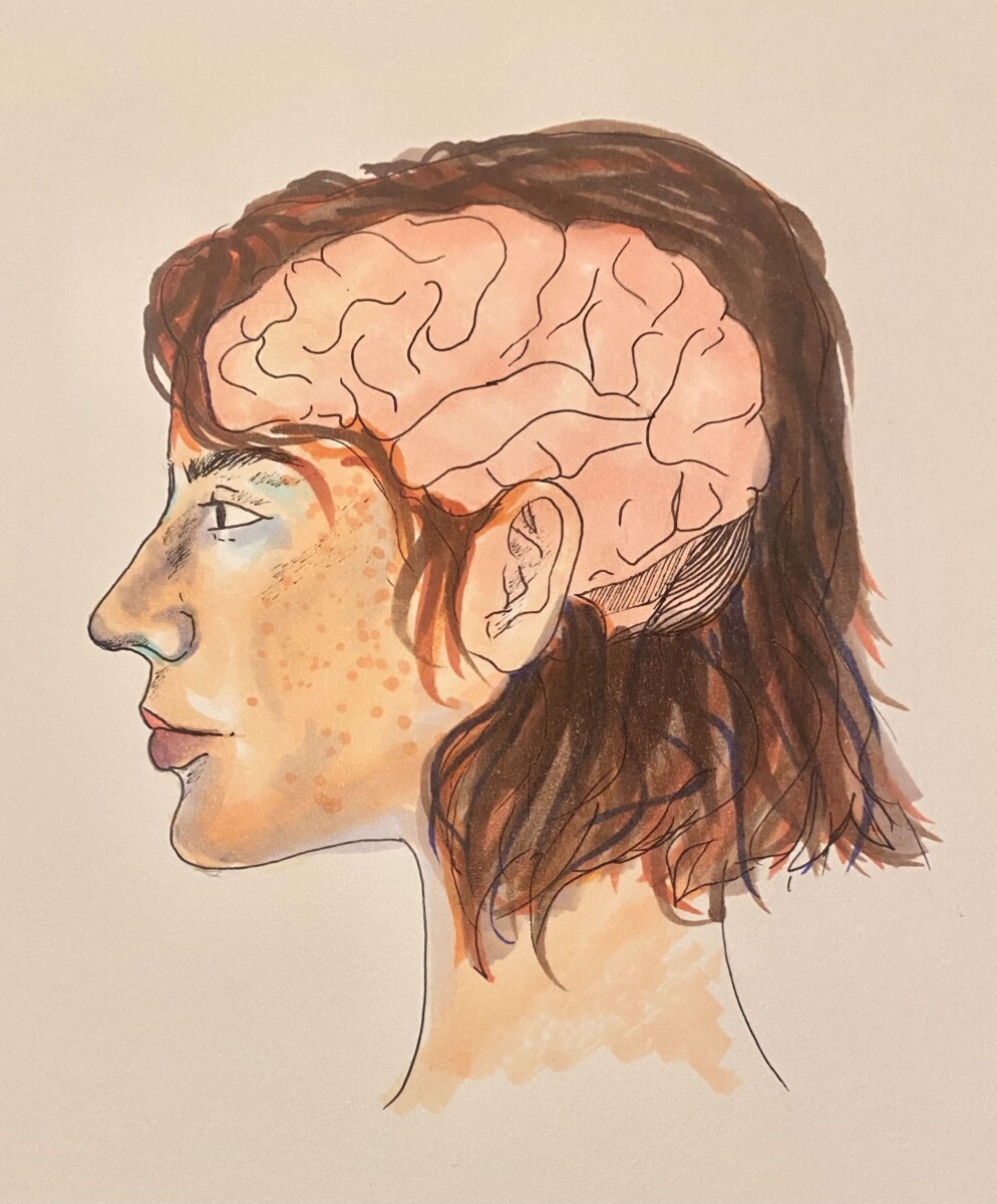
Learning a new language naturally occurs during the sensitive or critical periods when children have access to language exposure. During these specific time periods, the brain is able to rapidly learn a language due to cognitive processing capabilities and neural plasticity. Neural plasticity involves the brain modulating its functions and connections to accommodate newly acquired information, such as exposure to a new language. The neural plasticity in children’s frontotemporal systems allows for a large variety of languages to be acquired, and it has been demonstrated that there are significant similarities in language acquisition regardless of the modality or style. In particular, deaf children that have access to sign language during the sensitive time period acquire sign language using similar neural mechanisms to that of hearing children who use spoken language [1]. Access to language exposure during sensitive time periods is essential for long-term language proficiency. In cases with deaf children, research has shown that individuals with late exposure to sign language have less activation of the left frontotemporal pathways used for language comprehension [2]. The brain attempts to compensate for this by relying on other neural pathways. Typically, indications of strong right-side activity within the brain implies a lack of the left-side neural connections which form in early youth. However, even though the brain attempts to compensate for the lack of adequate exposure, there are still significant delays in language comprehension and proficiency [2]. Since language is a fundamental aspect of our social interactions, it is important that babies are screened for hearing impairments as screening allows caregivers to adjust their approach to ensure that the babies have adequate exposure to language. 
 

LangAcq_JeeyaSharma_3

Language acquisition that occurs in early life involves left hemisphere specialization [1]. Two important areas in the left hemisphere are Wernicke’s area and Broca’s area. Wernicke’s area refers to the left posterior superior temporal gyrus and the supramarginal gyrus, which are in front of the occipital lobe at the back of the brain, and Wernicke’s area is routinely referenced in discussions about the comprehension of language [3]. Recent research has supported that comprehension of key aspects of a first language is possible due to significant left temporal and inferior parietal lobe involvement, which demonstrates that speech comprehension is not isolated to Wernicke’s area. The left temporal and parietal lobes are located between the left frontal lobe and the left occipital lobe at the back of the brain, and their involvement indicates a more extensive language comprehension network. Additionally, Wernicke’s area appears to play a role in speech production [3]. Another area of the brain involved with language is the Broca’s area, which is located in the left inferior frontal gyrus, which is at the bottom of the frontal lobe [4]. It is traditionally defined as the key area involved with speech production, and modern research has demonstrated that the Broca’s area plays a vital role in sending information across the broader speech production neural networks. This contradicts earlier ideas that speech production is isolated to the Broca’s area [4]. Both of these findings demonstrate the large neural interactions between different areas of the left hemisphere that are responsible for language, and these interactions allow for language acquisition in early life to be successful long term. 


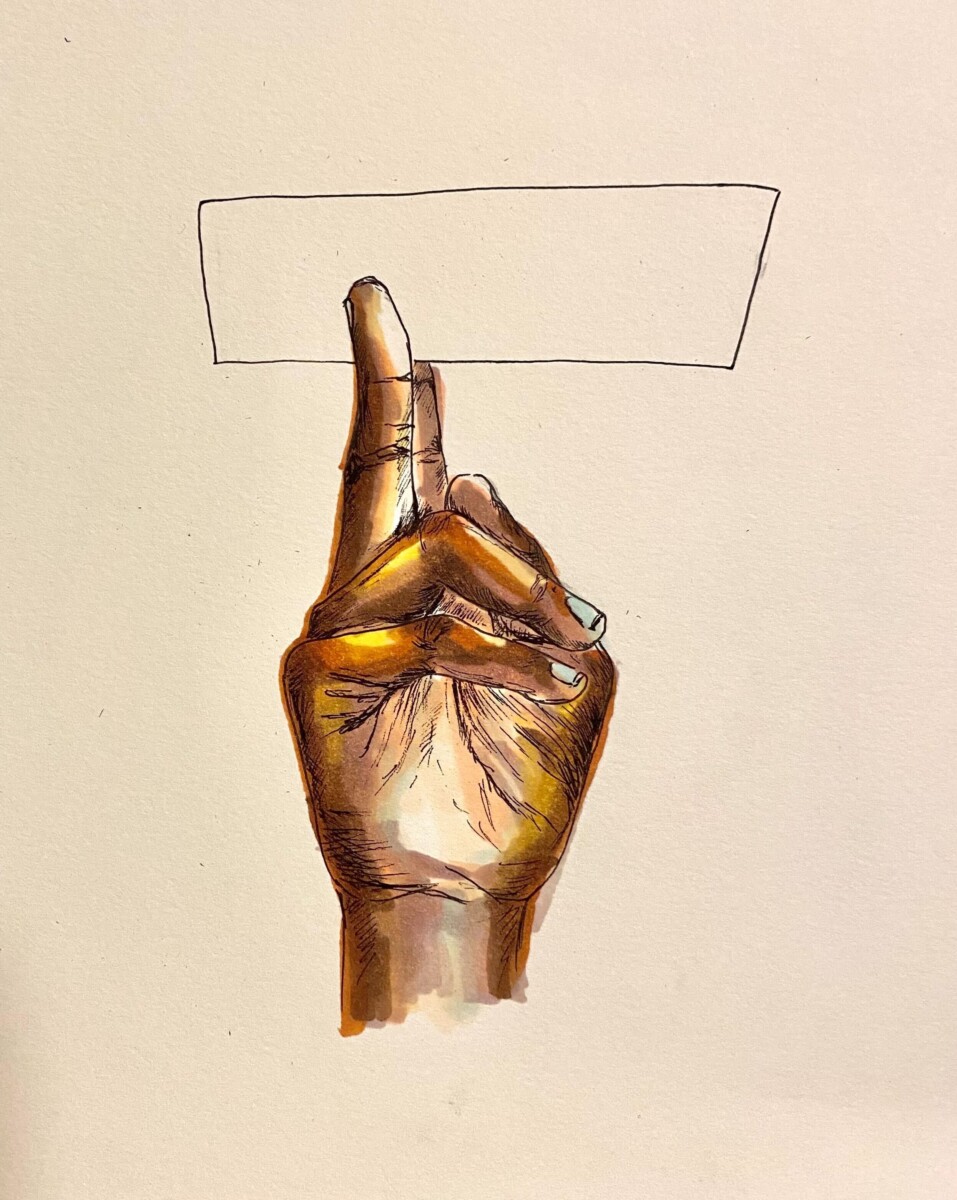
The acquisition of a second language is increasingly valuable in the modern world. Bilingualism is also beneficial on an individual level because of the associated cross-domain transfer and cognitive control observed throughout the lifespan [5]. When compared to monolingual individuals, bilingual individuals may experience improvements in non-linguistic general and executive functions, such as switching between tasks and selective monitoring [5]. The sensitive period for language learning also has implications on second language acquisition, but recent data suggests that the sensitive period appears to extend until adolescence for second language acquisition [6]. Additionally, while acquiring a first language after the sensitive period has significant consequences on socialization and cognitive development, the consequences associated with missing the sensitive period for second language acquisition are not as substantial. Limited proficiency in a second language can harm communication efforts within that language, but the stakes are lower for the second language in the regard that it does not interfere with typical development throughout childhood.
 

LangAcq_JeeyaSharma_2

While individuals that acquire a second language beyond the sensitive period have more limited proficiency long-term, older language learners may make significant gains in the short-term. Second language acquisition later in life may come with specific challenges, but the brain retains mechanisms of neuroplasticity that allows that acquisition to occur [5]. The first language can also interfere with second language acquisition, and when specific sounds are unfamiliar in the first language, it is especially difficult for individuals to make the necessary distinctions in the second language [5]. Despite those challenges, adults may be able to apply knowledge from the first language to the second language if there is a significant amount of overlap between the languages. In fact, recent research suggests that adults even outperform children during the short-term when they are learning a second language with similar materials [6]. However, children have long-term advantages in second language acquisition. Data has shown that there is a rapid decline in second language learning ability at around seventeen to eighteen years old, specifically in the ability to learn and comprehend grammar. It is currently not clear exactly why this occurs, but it may be due to changes in neural maturation and plasticity, increased interference from the first language, or cultural and social factors, including the common transition towards a career occurring in late adolescence. [6]. Ultimately, the findings of this research contradicted earlier claims that the sharp decline in language learning capabilities occurs before adolescence, and this also offers more favorable implications for the acquisition of a second language in late adolescence and early adulthood. 


Language acquisition within the sensitive period is a significant factor in typical childhood development; therefore, it is paramount that children receive adequate language exposure for lifelong language proficiency. Additionally, beyond this time period, the brain acquires language through the use of alternative mechanisms, which include right hemisphere involvement. The use of alternative mechanisms has a wide range of implications for second language learners, but data also suggests that second language acquisition later in life is still achievable. These findings provide valuable insight into human social behavior and communication mechanisms throughout the lifespan. 

Polyglot or Noob? Read More »

Love on the Brain

by Manju Karthikeyan

art by Danielle Mather

Love_DanielleM_MAIN

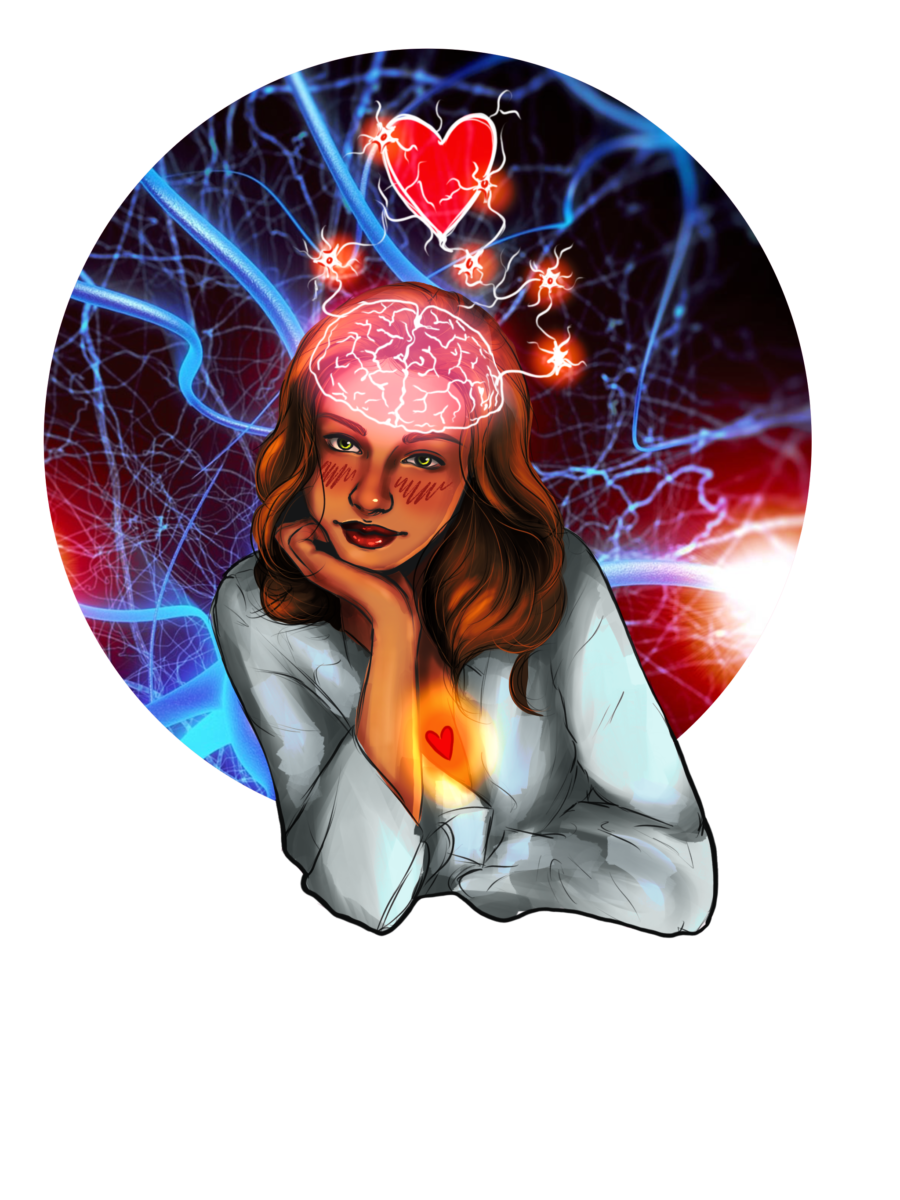
We all know that feeling… 

Your special someone walks into the room and before you know it, all ration is thrown out the window. Your heart races, your cheeks flush, and a swarm of butterflies start bursting in your stomach. 

But why is that? Why do our bodies and our brains fall into a frenzy in the face of attraction? And what is the effect of love, or rather, the chemistry of love, on our brains?


Topic 1: the immediate reactions, ANS, brain regions, etc.

The initial feeling of desire and attraction starts in a region of the brain known as the medial prefrontal cortex (MPFC). This region is like Tinder, in that it will immediately swipe right or left on what’s in front of you. The MPFC has the ability to instantaneously evaluate something as minute as facial appearance, making lasting judgments from a short conversation and dictating the brain’s decision-making process from there [1]. 

Once the medial prefrontal cortex (MPFC) sees something it likes, the rest of the body is alerted. The central nervous system (CNS) in general plays a crucial role in responding to what is desirable in a person. This is where those traditional symptoms of the “love bug” come in; sweating, blushing, and nausea. By intervening with the autonomic nervous system (ANS), the CNS will send a variety of signals that instigate your heart to elevate blood pressure, alert your pupils to dilate, and elicit parasympathetic responses to sexual desire as a whole [2, 3]. 

The prefrontal cortex isn’t the only player when it comes to love. The limbic system, responsible for mediating primal attraction and behaviors, is also activated in response to attraction in humans. In particular, the hypothalamus, the hippocampus, the caudate nucleus, the anterior cingulate cortex, and the ventral tegmental area (VTA) become active [4, 5]. 

The VTA is crucial to hardwiring the brain’s reward system and dopamine production. When we see something we are attracted to, the VTA is activated in the same way as if it were given a reward, reacting to love like a drug [6]. Moreover, the synaptic firings within the VTA, paired with the influx in dopamine that occurs, mimic neurological patterns similar to cocaine rushes in addiction circuits [7]. Thus, acting “high” when we’re in love is not that far-fetched of an idea. 

However, love, like the brain, has many grey areas. When considering love as a spectrum, from platonic relationships to lust, the science of attraction becomes far more complicated: a complex mix of neurotransmitters, hormones, and other chemical signals that influence behavior.


Topic 2: Role of neurotransmitters; love lasting vs. attraction and lust

At the basis of attachment is oxytocin– often referred to as the love hormone. Coupled with another hormone, vasopressin, oxytocin is responsible for the formation of romance and pair bonding– especially in the beginning stages of a relationship [8, 9]. Oxytocin and vasopressin receptors are abundant in regions like the hypothalamus, and will often interact with the dopamine reward system in the brain [8]. Levels of these hormones vary as you move through the different stages of love. Lust, given the primary focus on arousal and reproductive efforts, is more readily associated with activating sex hormones like testosterone and estrogen than with neurotransmitters [10]. 

However, the transition from lust to romantic attachment tends to reflect deficits in testosterone levels for men [11, 12]. When lust transitions into attachment, oxytocin and vasopressin begin to dominate. This differs from attraction, the intermediary stage between lust and attachment, where the brain’s behavior is particularly centered around rewards. As previously mentioned, dopamine reaches peak levels to provide a foundation for love’s reward-centric influences. Norepinephrine is also involved in this euphoria, which is why we sometimes can’t eat or sleep when we’re in love [10]. 


Topic 3: Love and attraction impairing our judgment/sense of obsession; anti-love brain technology + potential conclusion

Given this, it is evident that the neurotransmitters, hormones, and chemical signals induced during attraction alter our neurological state. But to what extent? 

One possibility is that love attacks the brain like an addiction. It has been noted that attachment-oriented pair-bonding mechanisms and chemical sequences often overlap with reward-learning and addiction mechanisms of the brain [13, 14]. Scientific literature further emphasizes that one can indeed get addicted to love, and that to be in love with someone is much like being addicted to them [13]. While there are differences in oxytocin levels between romantic love and drug addiction, dopamine reward patterns are quite similar [15]. 

Thus, the ability to get addicted to love is not improbable, and the experience of love in the brain can be overwhelming. If addiction or the consumption of drugs impairs our judgment, and love mimics those effects through a similar addiction-prone mechanism, how does love affect our decision-making ability, our social cognition, or our self-control?

A proposed approach to combating a love addiction is time. While the early stages of romantic love are similar to patterns of addiction, these symptoms diminish as the relationship progresses [15]. As futuristic as they sound, we can harness these patterns of addiction to create anti-love technologies to induce chemical breakups. 

With lust, there are a plethora of drug interventions through antidepressants and androgen blockers that inhibit the release of hormones like testosterone [13]. However, altering one’s sense of attraction and attachment is incredibly subjective, with many raising ethical questions of potentially dictating one’s ability to love. Nevertheless, there have been some MDMA drug trials to induce love and ecstasy in patients with PTSD. Similarly, SSRI interventions (involving the serotonin receptor) are known to cause emotional blunting, detachment, and an overall lack of romantic stimulation as a treatment for obsessive-compulsive disorder (OCD) [16, 17]. Additionally, drugs that stimulate love have been used to treat depressed patients [13, 16, 17]. However, the credibility, accessibility, and safety of these chemical breakups are controversial and yet to be determined. 


Nevertheless, this highlights the development of humans to love and be loved, creating a neurological dependency on affection. Love truly has an effect on the brain, mediated by a variety of hormones, neurotransmitters, and brain activations. So the next time you feel your heart racing, cheeks flushing, and butterflies bursting in your stomach from talking to that special someone, ask yourself: is this me, or my brain on love?


Love on the Brain Read More »

Synesthesia is Green, Synesthesia Tastes Like Oranges

by Sonali Poobalasingham

art by Kate Richardson

Synesthesia_KateRichar_MAIN

What do Billie Eilish, Pharrel Williams, Kanye West, Duke Ellington, and Stevie Wonder all have in common? Besides being accomplished musicians and singers, they are all synesthetes, or individuals with at least one form of synesthesia [1]. Synesthesia is formally defined as the phenomenon in which “stimulation of one [sensory] modality simultaneously produces sensation in a different modality” [2]. In other words, senses may blend together, leading to experiences such as experiencing music as color or associating certain words with a particular taste. While there are numerous forms of synesthesia, all cases share three defining characteristics: 1) a crossover between two or more of the five senses, 2) inducer-specificity (such as the letter “S” always being paired with the color pink), and 3) the ability to provide detailed accounts of synesthetic experiences [3]. In this article, we will start with hypotheses about the origins of synesthetic experiences and then explore recent research on grapheme synesthesia. Finally, we will investigate attempts to use hallucinogens to induce synesthetic perceptions. 


Synesthesia may be more common than you think – in infants, at least. A scientific review of synesthesia suggests that everyone is actually born a synesthete, challenging the widely-held assumption that synesthesia is a phenomenon that affects only a select few individuals. One hypothesis posits that the root cause of synesthesia in adults is abnormal synaptic pruning, or the process by which communication between brain cells is cut due to lack of use [4]. The human brain is composed of billions of brain cells called neurons. The synapse is the part of a neuron that connects with other neurons, allowing it to communicate with neighboring neurons. Infants have many synapses – the most they will ever have in their life, in fact! Because of these connections, infants’ five senses are very highly attuned, which induces synesthesia. Synaptic pruning is the process by which unneeded synapses between neurons are removed, leaving behind only the most efficient and useful connections to be carried into adulthood. If this seems abstract, picture the way a skilled gardener turns a shapeless hedge into a beautiful hedge sculpture just by cutting out the parts of the bush that were overgrown. This is something the human brain has the power to do to itself! Synaptic pruning in infanthood optimizes neural networks such that most do not experience synesthesia in adulthood. Pruning is a highly controlled process, but mistakes occasionally occur. This incomplete pruning hypothesis explains that an “overabundance of [neuronal] connections” caused by incomplete synaptic pruning allows different brain areas to remain cross-linked when their connections should have been severed [5]. This linkage, in turn, allows different sensory regions of the brain to be tied together, leading to the synesthetic perceptual experience in which certain senses are inextricably linked. 


To test this hypothesis, researchers compared levels of connectivity between brain areas in adults with and without synesthesia. Indeed, this study showed that synesthetes had considerably more connectivity – that is, more neural pathways connecting various brain areas – than individuals without synesthesia [5]. Additional evidence shows that hearing spoken words triggers both the auditory and visual cortices in infants, but this effect diminishes once the child reaches around three years of age [6]. This further supports the explanation that synesthetic experiences are produced by an excess of neuronal connections in the brain.


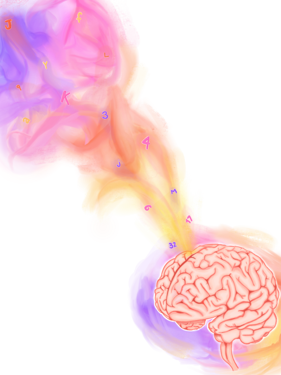
Now, let’s examine a specific subset of synesthesia called grapheme-color synesthesia. Of the numerous ways synesthesia can display itself, grapheme-color synesthesia is one of the most common presentations and is the best studied. Grapheme-color synesthesia occurs when the processing of numbers and letters becomes cross-wired with perception of color, leading to individuals associating colors with numbers and letters. These colors may appear over the number or letter being viewed, or the color may present itself “in the mind’s eye” [7]. The cross-activation theory, which proposes that many brain areas may fire together in response to a single stimulus, may explain the occurrence of grapheme-color synesthesia. In the brain, the visual word form area (VWFA) lies next to an area that processes color, called hV4. Proponents of the cross-activation theory believe that neurons in hV4 fire synchronously with neurons in the VWFA in grapheme-color synesthesia, leading to the experience of seeing colors when viewing numbers, letters, and words [8][9].  

Synesthesia_KateRichar_side1

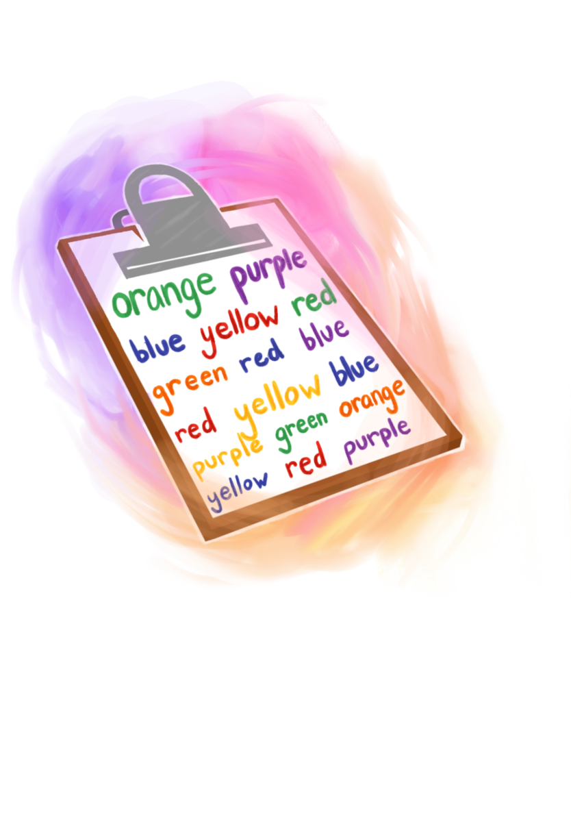
One way to study grapheme-color synesthesia is by using a form of the Stroop Test. The Stroop Test is a classic, well-documented psychological phenomenon: when instructed to read the word “blue,” most people can do so quickly and easily. However, when told to read the word “blue” when it is typed in a non-blue font, people often stutter, falter, and take longer to respond due to the conflicting word and text color [10]. Studies investigating grapheme-color synesthesia utilize the Stroop Test, but instead of changing the colors of words, the printed letter or number is made to differ from each individual’s synesthetic color of the character. If the printed character matches the synesthete’s color perception, the trial is called congruent; conversely, if the printed character does not match the synesthetic’s color perception, the trial is called incongruent [10]. For example, if the individual consistently saw the color blue while viewing the letter “Q”, a modified Stroop trial may involve displaying “Q” in the color blue first (a congruent trial) followed by “Q” being displayed in red (an incongruent trial). When synesthetic individuals were timed on how long it took to respond, they took significantly longer to respond in the incongruent trials compared to the congruent trials. Just as reading a presented word is the default response in classic Stroop trials, the default response in the modified Stroop trials is for a synesthetic individual to view the presented digit in the color that matches their synesthetic perception. In other words, the modified Stroop trials support the notion that individuals with grapheme-color synesthesia have no control over their synesthetic perceptions; their synesthetic experiences are as natural and involuntary as blinking. 


Another study on grapheme-color synesthetes sought to examine when exactly  synesthetic perceptions occur in the perceptual processing stages. Researchers used a congruence-incongruence model similar to the one used in the modified Stroop trials and combined it with a simple search task. In each trial, synesthetic participants were asked to find a letter when it is overlaid on a colored background. In congruent trials, the participant’s perceived color of the letter matched the background color; in incongruent trials, the participant’s perceived color of the letter did not match the background color [11]. For example, a congruent search trial for someone who associates “Q” with the color blue would consist of “Q” being displayed with a blue background, while an incongruent trial would consist of “Q” being displayed with any non-blue background. Across these search trials, synesthetes were able to identify the letter faster in the incongruent trials compared to in the congruent trials [11]. While this may seem like an obvious conclusion, it has fascinating implications. If synesthetic perception occurred in a later processing stage, synesthetes would quickly and easily be able to find a letter in a congruent trial before the perception of color appeared, because there would be a period of time in which the letter’s perceived color did not match the background, making it easily detectable. In order for background color to interfere with one’s ability to correctly identify a letter, the synesthetic perception of color (like seeing the letter “Q” as blue) must occur in an extremely early processing stage [11]. 

Synesthesia_KateRichar_side2

Interestingly, synesthesia-like experiences may also be temporarily inducible via psychoactive drugs. A controlled experimental study performed in 2013 showed that administration of lysergic acid diethylamide (commonly known as LSD) can induce synesthesia-like experiences in individuals without synesthesia, particularly perceptions of the grapheme-color and sound-color varieties [12]. However, these temporarily induced perceptions lack consistency and inducer-specificity; the links between graphemes, colors, and sounds are not reliably fixed. Consistency and inducer-specificity are important hallmarks of natural synesthesia that are lacking in LSD-induced perceptions. Nonetheless, the possibility of hallucinogens leading to synesthetic-like perceptions warrants more studies to learn more about how LSD causes synesthetic-like experiences.  


In summary, synesthesia is a very fascinating neurological condition. Current research suggests that synesthesia is universal during infancy, and that the neural connections responsible for synesthetic experiences are ordinarily severed via synaptic pruning. The incomplete pruning hypothesis proposes that synesthesia in adults is caused by incomplete synaptic pruning that has left unrelated neural connections intact, as seen in grapheme-color synesthetes demonstrating robust connections between the VWFA and the hV4 brain areas. Hallucinogens such as LSD have been shown to temporarily induce synesthesia-like experiences in non-synesthetes. Studies utilizing a modified Stroop Test and search tasks in individuals with grapheme-color synesthesia support the notion that synesthetic perceptions occur in a very early processing stage. While synesthesia has gained more attention in recent decades, there is still a lot undiscovered about it that merits future scientific study. After all, what better demonstrates the rich human experience if not examining the delicate, beautiful interplay between the human brain and perception of the world around us?

Synesthesia is Green, Synesthesia Tastes Like Oranges Read More »

The Rampant Disease We Call Social Discrimination

by Chloe Helsens

art by Owen Helsens & Kayla Barry

SocialDis_KaylaBar_MAIN
  1. What is social discrimination? 

            Imagine a disease, one that humans have created, is ravaging our population, and we are fallaciously withholding the cure from those who are suffering most. In a sense, that is exactly what social discrimination is: we made it, and although it’s preventable, we turned a social construct into a tangible reality within each victim’s body. Social discrimination is “the differential treatment of individuals based on their ethnicity, cultural background, social class, educational attainment, or other sociocultural distinctions” [1]. Social discrimination is rampant throughout societies around the world and is widely recognized as a type of psychological stressor, being detrimental to the mental state. Discrimination has had — and continues to have – immeasurably damaging effects on people all over the globe. Recent studies have investigated how the distress of social discrimination leads to negative physical and mental health outcomes. We will examine how a discriminatory society has forced its way into our physiological functions.

SocialDis_KaylaBar_side2
  • How does social discrimination change our brains?

There are hundreds of types of discrimination that remain rampant throughout societies for centuries, yet only contemporary scientific studies have begun to recognize the severity of social discrimination’s effects on the brain. For instance, a recent 2017 study examined seventy-four adults from traditionally marginalized communities where all participants completed a self-report regarding their exposure to discrimination. They were assessed through a functional magnetic resonance imaging (fMRI) scan, which measures blood flow in the brain. The fMRI scan measured participants’ amygdala activity, a region in the brain associated with emotion, and functional connectivity, which measures the interactions between two regions of the brain. The results found that individuals reporting greater incidences of discrimination were correlated with having greater amygdala activity. They also discovered increased connectivity between the amygdala and brain regions associated with the salience network, which are multiple regions in the brain that tell us which stimuli we should pay attention to [2]. The results were eye-opening because greater amygdala activity in the brain is associated with higher levels of chronic stress, and greater connectivity between the two regions “has been reported in individuals with PTSD and single-episode depression”  [3]. In summary, adults from traditionally marginalized communities had neural activity reflective of extremely stressed individuals, which can lead to higher risks for chronic stress and depression. This sheds light on the evermore apparent links between social discrimination and negative neurophysiological outcomes. 

Another study examining how US college students’ mental health is affected by social discrimination also found distressing results. Using data collected from the National College Health Assessment (NCHA), researchers surveyed around 417,000 college students from a myriad of universities across the US. They found that out of the 7.9% of students that experienced social discrimination in some form, there was a 37% increase in mental health symptoms and a 94% rise in the number of mental health diagnoses compared to students who reported no discriminatory experiences [4]. They also found other alarming statistics; for example, cisgender men who experienced discrimination were 210% more likely to consider suicide, 900% more likely to attempt suicide, and over 1000% more likely to self-diagnose with schizophrenia compared to cisgender men who had not experienced discrimination. High amounts of discriminatory experiences were reported most among Hispanic/Latino, African-American, Asian, and multiracial students, although Indigenous students had the highest association with poor mental health outcomes [4].  

SocialDis_KaylaBar_side1
  • Allostasis as a result of continual discriminatory encounters 

Now that we see the abhorrent impacts that discrimination has on mental health, it is no surprise that these effects can also manifest as physiological functions. But how do we get from discrimination to disease? Only recently have scientists discovered through what means this can occur; allostatic load, or the body’s response to the cumulative burden on itself because of chronic stress [5]. One example of the effects of allostatic load on the body is shown by a 2014 study conducting a meta-analysis of the physiological impacts of racial discrimination. They found that allostatic load, the culmination of life stress and its effects on the body, can exacerbate the effects of discrimination that the body feels by leading to pathophysiology, or thwarting how the body normally functions. Symptoms can include chronic stress, metabolism changes, and increased disease vulnerability. Essentially, the greater the chronic stress associated with social discrimination, the greater the cumulative impact on their health.

Racial discrimination can also lead to the over-activation of the hypothalamic-pituitary-adrenal (HPA) axis, which is a feedback system in the body where different hormone pathways communicate and interact with each other; principally, it acts as our body’s primary stress response pathway [6]. This over-activation of the HPA axis produces increased levels of cortisol, a hormone commonly released as a response to stress. Chronic release of cortisol can cause “metabolic changes and effects on the immune system as well as behavioral alterations,” which connects physiological effects, such as “mood changes and cognitive impairment,” to discrimination [7]. In conclusion, social discrimination can lead to an allostatic overload, acting as the mechanism for discrimination to convert itself into diseases within the body. 

Not only does discrimination affect our immune system and behavior, but it also activates a separate axis in the body that is triggered by stress, called the sympathetic-adrenal-medullary axis (SAM), that changes our cardiovascular function by increasing blood pressure, heart rate, etc [8]. The over-activation of these pathways consequently becomes a catalyst for heart conditions such as cardiovascular disease. In congruence with these findings, traditionally marginalized groups in the US, such as African Americans and Hispanics, had “chronic cardiovascular, inflammatory, and metabolic risk factors,” likely because of the discrimination they experience on a daily basis, according to Berger [9].

More recent research uncovered the fact that discrimination compromises brain matter integrity due to constant stress states. The study was conducted at Emory University with two groups consisting of both Black and White females, and investigated links between incidences of medical disorders, racial discrimination, and white matter integrity [10]. White matter enables information to be transmitted between different structures within the brain [11]. The scientists found that white matter can act as a bridge between discrimination and physiological health through chronic inflammation. Chronic inflammation damages the connectivity of white matter, which then translates to diseases such as hypertension, cardiovascular disease, and arthritis. Dr. Fani, along with her fellow researchers, found a pathway in which racist experiences may increase the risk for health problems via effects on select stress-sensitive brain pathways. They also linked these changes to “risk for negative health outcomes, possibly influencing regulatory behaviors.” “Now we can see that these changes may enhance the risk for negative health outcomes, possibly by influencing regulatory behaviors” [10]. Altogether, these studies expose the pathways in which discrimination harms marginalized groups, and how it thrashes its way into our physiological functions. 

How much longer are we going to tolerate social discrimination within our societies? Why is it still acceptable? We continually say, in theory, we are advocates for inclusivity and social justice, yet shy away from condemning our peers, friends, or family members’ discriminatory remarks. To what extent will countless bodies pay for a sickness our society instituted? Discriminatory experiences accumulate in our bodies, eliciting both psychological and physiological consequences, while also altering stress load, white matter integrity, and even our risk for diseases. Social discrimination continues to seep its way into all facets of our world, while many of us passively watch as it rots our brains and bodies. Instead of watching as societal discrimination manifests itself into mental health disorders, diseases, and other horrifying effects, we should finally recognize and enact preventative measures against the overwhelming mental and physical burden discrimination poisons us with.

The Rampant Disease We Call Social Discrimination Read More »