by Sonali Poobalasingham
art by Kate Richardson

What do Billie Eilish, Pharrel Williams, Kanye West, Duke Ellington, and Stevie Wonder all have in common? Besides being accomplished musicians and singers, they are all synesthetes, or individuals with at least one form of synesthesia [1]. Synesthesia is formally defined as the phenomenon in which “stimulation of one [sensory] modality simultaneously produces sensation in a different modality” [2]. In other words, senses may blend together, leading to experiences such as experiencing music as color or associating certain words with a particular taste. While there are numerous forms of synesthesia, all cases share three defining characteristics: 1) a crossover between two or more of the five senses, 2) inducer-specificity (such as the letter “S” always being paired with the color pink), and 3) the ability to provide detailed accounts of synesthetic experiences [3]. In this article, we will start with hypotheses about the origins of synesthetic experiences and then explore recent research on grapheme synesthesia. Finally, we will investigate attempts to use hallucinogens to induce synesthetic perceptions.
Synesthesia may be more common than you think – in infants, at least. A scientific review of synesthesia suggests that everyone is actually born a synesthete, challenging the widely-held assumption that synesthesia is a phenomenon that affects only a select few individuals. One hypothesis posits that the root cause of synesthesia in adults is abnormal synaptic pruning, or the process by which communication between brain cells is cut due to lack of use [4]. The human brain is composed of billions of brain cells called neurons. The synapse is the part of a neuron that connects with other neurons, allowing it to communicate with neighboring neurons. Infants have many synapses – the most they will ever have in their life, in fact! Because of these connections, infants’ five senses are very highly attuned, which induces synesthesia. Synaptic pruning is the process by which unneeded synapses between neurons are removed, leaving behind only the most efficient and useful connections to be carried into adulthood. If this seems abstract, picture the way a skilled gardener turns a shapeless hedge into a beautiful hedge sculpture just by cutting out the parts of the bush that were overgrown. This is something the human brain has the power to do to itself! Synaptic pruning in infanthood optimizes neural networks such that most do not experience synesthesia in adulthood. Pruning is a highly controlled process, but mistakes occasionally occur. This incomplete pruning hypothesis explains that an “overabundance of [neuronal] connections” caused by incomplete synaptic pruning allows different brain areas to remain cross-linked when their connections should have been severed [5]. This linkage, in turn, allows different sensory regions of the brain to be tied together, leading to the synesthetic perceptual experience in which certain senses are inextricably linked.
To test this hypothesis, researchers compared levels of connectivity between brain areas in adults with and without synesthesia. Indeed, this study showed that synesthetes had considerably more connectivity – that is, more neural pathways connecting various brain areas – than individuals without synesthesia [5]. Additional evidence shows that hearing spoken words triggers both the auditory and visual cortices in infants, but this effect diminishes once the child reaches around three years of age [6]. This further supports the explanation that synesthetic experiences are produced by an excess of neuronal connections in the brain.
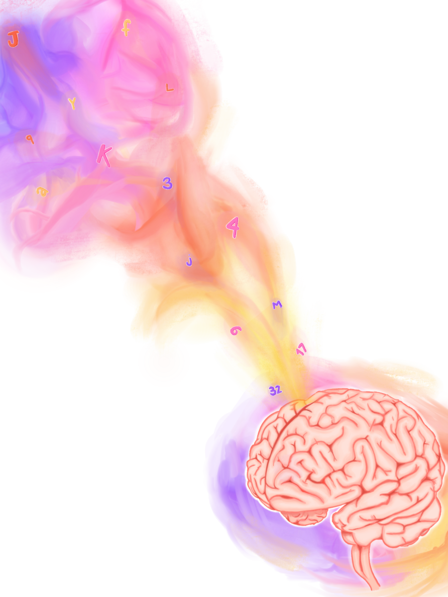
Now, let’s examine a specific subset of synesthesia called grapheme-color synesthesia. Of the numerous ways synesthesia can display itself, grapheme-color synesthesia is one of the most common presentations and is the best studied. Grapheme-color synesthesia occurs when the processing of numbers and letters becomes cross-wired with perception of color, leading to individuals associating colors with numbers and letters. These colors may appear over the number or letter being viewed, or the color may present itself “in the mind’s eye” [7]. The cross-activation theory, which proposes that many brain areas may fire together in response to a single stimulus, may explain the occurrence of grapheme-color synesthesia. In the brain, the visual word form area (VWFA) lies next to an area that processes color, called hV4. Proponents of the cross-activation theory believe that neurons in hV4 fire synchronously with neurons in the VWFA in grapheme-color synesthesia, leading to the experience of seeing colors when viewing numbers, letters, and words [8][9].

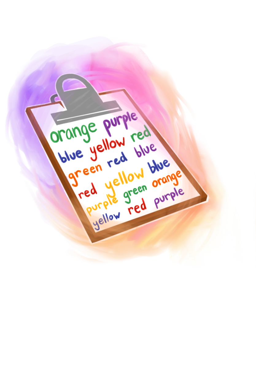
One way to study grapheme-color synesthesia is by using a form of the Stroop Test. The Stroop Test is a classic, well-documented psychological phenomenon: when instructed to read the word “blue,” most people can do so quickly and easily. However, when told to read the word “blue” when it is typed in a non-blue font, people often stutter, falter, and take longer to respond due to the conflicting word and text color [10]. Studies investigating grapheme-color synesthesia utilize the Stroop Test, but instead of changing the colors of words, the printed letter or number is made to differ from each individual’s synesthetic color of the character. If the printed character matches the synesthete’s color perception, the trial is called congruent; conversely, if the printed character does not match the synesthetic’s color perception, the trial is called incongruent [10]. For example, if the individual consistently saw the color blue while viewing the letter “Q”, a modified Stroop trial may involve displaying “Q” in the color blue first (a congruent trial) followed by “Q” being displayed in red (an incongruent trial). When synesthetic individuals were timed on how long it took to respond, they took significantly longer to respond in the incongruent trials compared to the congruent trials. Just as reading a presented word is the default response in classic Stroop trials, the default response in the modified Stroop trials is for a synesthetic individual to view the presented digit in the color that matches their synesthetic perception. In other words, the modified Stroop trials support the notion that individuals with grapheme-color synesthesia have no control over their synesthetic perceptions; their synesthetic experiences are as natural and involuntary as blinking.
Another study on grapheme-color synesthetes sought to examine when exactly synesthetic perceptions occur in the perceptual processing stages. Researchers used a congruence-incongruence model similar to the one used in the modified Stroop trials and combined it with a simple search task. In each trial, synesthetic participants were asked to find a letter when it is overlaid on a colored background. In congruent trials, the participant’s perceived color of the letter matched the background color; in incongruent trials, the participant’s perceived color of the letter did not match the background color [11]. For example, a congruent search trial for someone who associates “Q” with the color blue would consist of “Q” being displayed with a blue background, while an incongruent trial would consist of “Q” being displayed with any non-blue background. Across these search trials, synesthetes were able to identify the letter faster in the incongruent trials compared to in the congruent trials [11]. While this may seem like an obvious conclusion, it has fascinating implications. If synesthetic perception occurred in a later processing stage, synesthetes would quickly and easily be able to find a letter in a congruent trial before the perception of color appeared, because there would be a period of time in which the letter’s perceived color did not match the background, making it easily detectable. In order for background color to interfere with one’s ability to correctly identify a letter, the synesthetic perception of color (like seeing the letter “Q” as blue) must occur in an extremely early processing stage [11].

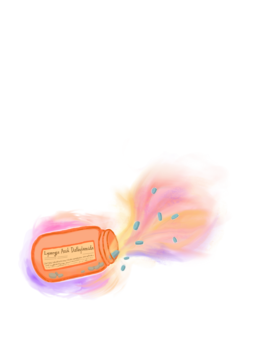
Interestingly, synesthesia-like experiences may also be temporarily inducible via psychoactive drugs. A controlled experimental study performed in 2013 showed that administration of lysergic acid diethylamide (commonly known as LSD) can induce synesthesia-like experiences in individuals without synesthesia, particularly perceptions of the grapheme-color and sound-color varieties [12]. However, these temporarily induced perceptions lack consistency and inducer-specificity; the links between graphemes, colors, and sounds are not reliably fixed. Consistency and inducer-specificity are important hallmarks of natural synesthesia that are lacking in LSD-induced perceptions. Nonetheless, the possibility of hallucinogens leading to synesthetic-like perceptions warrants more studies to learn more about how LSD causes synesthetic-like experiences.
In summary, synesthesia is a very fascinating neurological condition. Current research suggests that synesthesia is universal during infancy, and that the neural connections responsible for synesthetic experiences are ordinarily severed via synaptic pruning. The incomplete pruning hypothesis proposes that synesthesia in adults is caused by incomplete synaptic pruning that has left unrelated neural connections intact, as seen in grapheme-color synesthetes demonstrating robust connections between the VWFA and the hV4 brain areas. Hallucinogens such as LSD have been shown to temporarily induce synesthesia-like experiences in non-synesthetes. Studies utilizing a modified Stroop Test and search tasks in individuals with grapheme-color synesthesia support the notion that synesthetic perceptions occur in a very early processing stage. While synesthesia has gained more attention in recent decades, there is still a lot undiscovered about it that merits future scientific study. After all, what better demonstrates the rich human experience if not examining the delicate, beautiful interplay between the human brain and perception of the world around us?
