Category Archives: Uncategorized

The Bridge Between Recollection and Inception

Dear family and friends,

The priority I set for myself in coming to Paris (besides academics of course, duh!) is sightseeing. This is my first time in Paris, which means every turn I take is a new location I have yet to explore. Thus, even with the unwelcome addition of jet lag and travel-induced dehydration I have already visited several attractions . Notable mentions include the Eiffel Tower, the Galeries Lafayette, the Arc de Triomphe, and the Notre Dame Cathedral. However, the most exciting place I have visited is one you may not recognize – a bridge named Pont de Bir-Hakeim. While I know absolutely nothing about the walkway’s historical significance, I do know one very important detail. It is the bridge that Leonardo Dicaprio and Ellen Page march across in Christopher Nolan’s 2010 blockbuster film Inception. By now you have probably figured that when I say “exciting” it is almost entirely subjective. Nonetheless, the bridge is quite picturesque and presents a great view of the Eiffel Tower.


View of the Eiffel Tower from Pont de Bir-Hakeim


Pont de Bir-Hakeim

When I first saw the bridge I was a few hundred meters away. It immediately looked familiar to me; however, I could not place in my memory where I had seen it before. After several minutes of contemplation: poof! Memories flooded my mind of the scene in Inception and the joy I felt after watching the film ten times. I was so delighted that I moved the bridge to the top of my to-do list and visited it several days later with some friends. My successful recollection got me thinking – how could I recognize the bridge before remembering specific details about it? It turns out neuroscience has an answer!


Standing in Leo’s footsteps (Photo by Chandler Lichtefeld)

To start, recognition memory is formally split into two categories: recollection and familiarity. Both occur in response to a previously experienced stimulus e.g. an event, person, or object. Recollection describes a person’s ability to retrieve specific details about the previously experienced stimulus. Familiarity is one’s feeling that the stimulus was previously experienced, without retrieval of explicit details. To simplify, think of recollection as “remembering” and familiarity as “knowing.” Recently, a group of researchers set out to clarify the neural correlates of each recognition process. Led by Dr. Jeffrey Johnson at the University of Missouri, researchers used functional magnetic resonance imaging (fMRI) to measure the brain activity of 20 participants during a memory task (Johnson et al. 2013). This memory task consisted of two parts: an encoding phase and a retrieval phase. During the encoding phase, word stimuli were presented visually to the participants. Words denoted single objects such as tools, animals, and food. Participants memorized the words by either putting them in a sentence (sentence condition) or associating their physical manifestation with an outdoor scene (scene condition). Basically, the encoding task required participants to use different methods to (hopefully) remember word stimuli. Next, the retrieval phase tested the participants’ ability to remember the previously presented word stimuli. Here, old word stimuli from the encoding phase and new word stimuli were presented on a neutral background. During presentation of words, participants could answer in several ways. Answering “R” meant that the subject remembered details about the word i.e. they remembered the sentence they made or the scene with which the word was associated. Therefore, answering “R” indicated that the subject could “recollect” details about the previous word. If unable to remember details, participants answered based on their confidence that the word was old or new. For example, answering with ”confident old” indicated that the subject was only “familiar” with the word.

So… what did the results show?

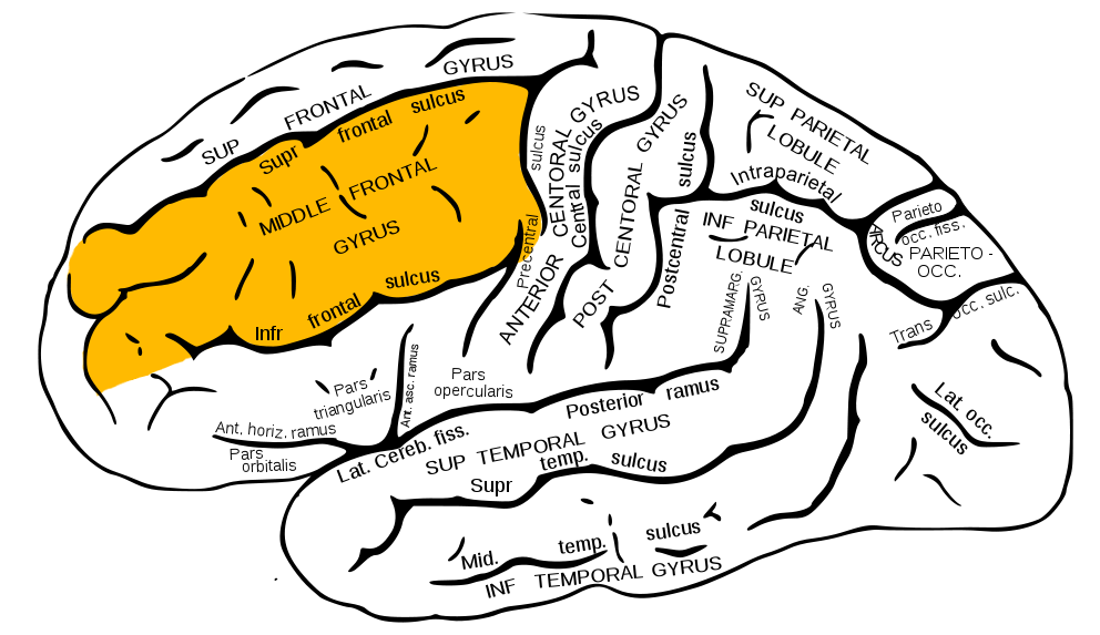
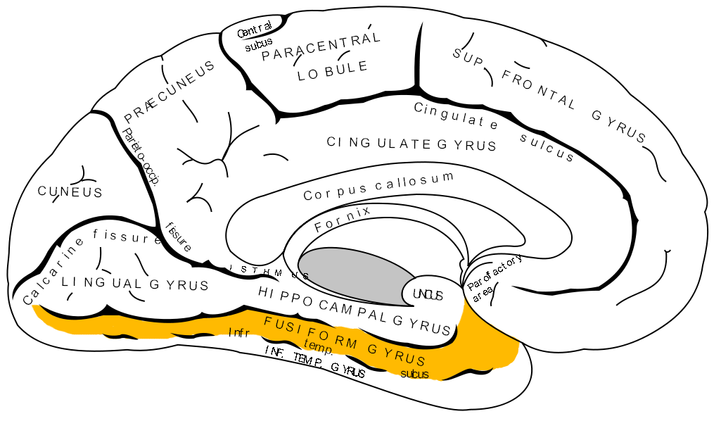
According to the imaging data, recollection-driven recognition activates different brain areas than familiarity-driven recognition. In other words, the mental processes behind recollection (remembering) are different than that of familiarity (knowing). Specifically, recollection (when participant sanswered “R”) activated the angular gyrus, left ventral parietal cortex, retrosplenial and posterior cingulate cortex, ventromedial PFC, bilateral hippocampus, and the bilateral posterior parahippocampal cortex. On the other hand, familiarity (when participants answered with “confident old”) activated the left intraparietal sulcus, precuneus, anterior cingulate, and dorsolateral PFC (Wow – that is a mouthful). Thus, according to this group’s rationale, it would seem that my initial recognition of Pont de Bir Hakeim was due to unique “familiarity” brain circuitry. As soon as I remembered details about the bridge, my “recollection” brain areas activated and brought forth memories of the movie’s bridge scene. Cool stuff, no?


Neural correlates of familiarity and recollection


Although the researchers failed to present some behavioral data due to too few trials, I thought that this study was well designed overall. They used copious background studies to support the rationale for their experimentation and produced results that clarify our current understanding of recognition-based memory. An interesting next step might be to examine latency time between familiarity and recollection in cases where one eventually remembers why a stimulus is familiar. Perhaps then I could understand why it took me a few minutes to recollect the bridge!


Until next time,

Christian


References

Johnson JD, Suzuki M, Rugg MD (2013) Recollection, familiarity, and content-sensitivity in lateral parietal cortex: a high-resolution fMRI study. Front Hum Neurosci 7:219.

All pictures were taken by myself and Chandler Lichtefeld (The picture of Leonardo DiCaprio and Ellen Page is a screenshot from my iTunes copy of Inception)

The brain activity figures were taken from the primary article by Johnson et al.

Making Memories One Sniff at a Time

Earlier this week we visited Le Grand Musée du Parfum, or for those of you who don’t speak French, the grand perfume museum (kind of easy to guess). Before arriving, I didn’t know what to expect besides that we would smell a whole lot of perfume. I was right, the building was filled with a variety of fragrances waiting to be sniffed, but this was not the only thing the museum contained. We walked through a maze of rooms displaying all sorts of information about perfume, starting with a historical journey of the origins from ancient Egypt to the Roman Empire and all the way to present time. Following the history was a sensory immersion exhibit (my personal favorite being a neuroscientist) that explained how our sense of smell works and contained olfactory games and fragrant riddles. Lastly, the museum had an exhibit dedicated to the art of the perfumer, where they had a collection of raw materials, natural and synthetic, most commonly used by perfumers. By the end of the museum my odor receptors were exhausted.

smelling one of the perfumer’s raw materials

My favorite room of the museum was the jardin des senteurs, or garden of the scents. We were told to walk up to these large white flowers, close ours eyes, breathe in the odor, try to guess what scent we were smelling and see if it triggered any memories. I did exactly this and took a big whiff of the first odor. Immediately I could recognize the smell of a campfire and a memory was triggered. I pictured myself sitting around a fire with my dad and sister and we were roasting marshmallows, an activity I love to do! I opened my eyes and was surprised and fascinated at the same time by this result. I quickly moved on to the next flower, closed my eyes, and sniffed. I was instantly at my grandma’s house on Christmas morning and an aroma floated through the air. It was cinnamon! I was reminded of the freshly baked cinnamon cookies we made around the holidays.

Jardin des Senteurs (garden of the scents)

Engrossed by this activity, I wondered if different areas of the brain were used when forming and retrieving memories of events in the presence and absence of strong odors. I did some googling and found a recent study that investigated the brain areas involved in episodic memory retrieval, or memories of a specific event, depending on the presence of an odor during encoding, the initial learning of new information (Galliot et al., 2013). Participants in the study consisted of thirteen female students between the ages of 20 and 23 (interestingly no males were used because olfactory abilities and brain regions can differ between genders). The experimental task consisted of two stages. In the first stage (encoding), 32 colored pictures of objects or animals were presented on a computer screen and participants were asked to determine if each picture contained more or less than three colors. This ensured the participants examined each picture carefully, but remained unaware the test was related to memory. During this task participants wore a mask with a valve that contained filter paper soaked in either water or vanillin, an olfactory stimulus usually considered pleasant. Half of the participants wore a mask with vanillin odor for the first 16 pictures and the other half wore the water filtered mask. The participants switched masks for the second 16 pictures.

types of memory

Two weeks later, the second stage (recognition) of the experiment was conducted. During this stage, each of the pictures used in the first stage (target) were presented simultaneously with a new picture (distractor). After the presentation of the two pictures, participants were instructed to press either a left or right button according to the side of the computer screen the participant believed was the target picture. For the duration of this task, participants were in an fMRI machine so that the investigators could record their brain activity. They found brain areas known to be strongly associated with episodic memory retrieval, the posterior hippocampal formations and the anterior thalamic nucleus, were activated whether or not an odor was presented in the first stage. However, they did find that learning in the presence of an odor causes activation of additional brain areas during the retrieval task. One of these areas was the orbitofrontal cortex and it has been previously described as the main site of secondary olfactory processing. They also found other areas in the frontal lobe of the brain, the superior, middle, and inferior frontal gyri (the bumps on the brain), were activated more during presentation of images encoded in the presence of the vanillin odor. However, the specific role in olfaction of these three brain areas remains unclear. I was very fascinated by the results that memories made in the presence of odors activated different brain regions during retrieval.

orbitofrontal cortex

The study also found that there was no significant difference between the number of correct responses of the target images between the pictures encoded with the presence of an odor and the pictures encoded without an odor. This finding made me wonder if the researchers had presented the odor during the retrieval stage of the experiment, would it increase the number of correct responses of the target images encoded with the vanillin odor? When I smelled the campfire and cinnamon odors, my memories were triggered instantly, so I would hypothesize if the participants smelled the vanillin during the recognition task, it would enhance their memory and would increase the number of correct target responses for the pictures encoded with the odor.

Now as I walk through the streets of Paris smelling the freshly baked breads and desserts, I will be reminded that the memories I form will cause different areas of my brain to be activated among retrieval.

Sources:

Galliot E, Comte A, Magnin E, Tatu L, Moulin T, Milot J (2013) Effects of an ambient odor on brain activations during episodic retrieval of objects. Brain Imaging and Behavior 7:213219.

Pictures 1 and 2 were taken by Dr. Kristen Frenzel

Picture 3: http://www.mindauthor.com/psychology/semantic-episodic-memory/

Picture 4:  https://commons.wikimedia.org/wiki/File:MRI_of_orbitofrontal_cortex.jpg

The Paris Brain without High-fructose corn syrup

Most people close to me know that nutrition plays a large role in my life, and I am very passionate about it. In the future, I would love to be able to have a job in which I can use my academic knowledge of neuroscience with my passion for nutrition to help improve others’ lives.

In America I feel like I constantly have to examine ingredient labels searching for chemicals that are unnecessary and harmful to my health. Often times food marketing is extremely misleading and without close examination, it is easy to fall trap to the commercialized mass production of lab created and modified food.

Arriving to Paris, I knew food is a large part of French culture, so I was interested to experience how the French viewed food and nutrition by living amongst their culture for the 5 weeks of this program.

Thus far, about 2 weeks into the program, I already see many clear distinctions between the two cultures. As I am sitting in a Parisian café writing this blog post, I cannot help but question whether it was just my imagination or if the food tasted fresher and cleaner.   At home, most people would view my diet as very restricted since I shy away from most breads, dairy items, any processed foods, and added sugars. I do this because I feel the way these items have been made is not beneficial to my overall health.   Since being in Paris, however, I feel fine eating some bread and cheese with my meals, common additions and cultural components of a French meal, as they taste fresher, cleaner and less processed than what one would find in a typical restaurant in America.

One of my many meals at this café in Paris

These observations along with my passion for nutrition and neuroscience, led me to want to delve further into specific components of food banned in France, as well as the rest of Europe, but commonly found in American food.

This led me to…High-Fructose Corn Syrup.

High-fructose corn syrup

The research article I found was conducted at Emory, which I did not realize until after I read it and thought it would be good to include!

Many previous studies have concluded that high-fructose corn syrup contributes to obesity through metabolic dysregulation, which is an umbrella term to describe the many processes in the body that are disrupted and ways obesity can impact our health. Previous studies have also examined the ways in which it can affect stress, which then can contribute to anxiety and depression. This study, conducted by Harrel et al (2015), aimed at looking at the implications high-fructose corn syrup had on our mental health, specifically in adolescents with developing brains. The researchers were looking at whether this sweetener had long-term implications on our response to stress, and to test this, they took rats and gave some of them a diet for 10 weeks with high fructose corn syrup and others a standard diet. All the rats were put under situations that induced stress for 12 days. The researchers then tested all the mice with situations to see behaviors associated with anxiety or depression. Essentially the researchers

Hypothalamus is the area in the brain in blue

found that with a high fructose diet, not only could they increase stress hormones like previous studies showed, but they could also induce anxiety and depressive behaviors, as well as induce changes in gene expression in the brain, specifically the area called the hypothalamus.

It is important to note that when replicated in adult rats, researchers found that the sweetener did not have an effect. Thus, fructose is affecting the adolescent developing brain on an intricate level and can lead to future poor mental health outcomes.

A strength that I liked of this article is that it tested adult rats, so that we could specifically see the danger is fructose consumption on a developing individuals brain. I would have liked to see a diet that contained high fructose corn syrup, along with other well-known super foods to see whether an exceptionally healthy diet could negate the harmful effects of high-fructose corn syrup. Not a limitation, but rather a question this article brought up for me, is replicating the exact study, but substituting high-fructose corn syrup for one of the many other supposedly harmful substances found in foods banned in Europe but not America like Stevia, food dyes, GMO’s and certain pesticides.

Feelings associated with Depression and poor mental health

Being a student in college, I am surrounded by many individuals close to me who confide about their personal experiences with anxiety and depression. The amount is astounding, and it should not be the norm. As someone interested in both nutrition and the brain, this leads me to question whether diet plays a role in this. Depression and anxiety are widespread problems. Having something commonly present in the food we consume that has been shown by this study to induce depression and anxiety cannot be beneficial to our mental health. Do Parisians have an advantage with not having to worry that a harmful chemical exists in their food?

For now and the rest of my time in Paris, I will enjoy eating without worry, especially all the baguettes made from fresh, local ingredients, cheese not modified in a lab, and dessert without high-fructose corn syrup. However, once back in the United States, I hope that the regulations soon catch up with the science, so that I do not have to worry that something harmful to my mental health is present in the food I put into my body.


Bibliography:

Harrell, C. S., Burgado, J., Kelly, S. D., Johnson, Z. P., & Neigh, G. N. (2015). High-fructose diet during periadolescent development increases depressive-like behavior and remodels the hypothalamic transcriptome in male rats. Psychoneuroendocrinology, 62, 252–264. http://doi.org/10.1016/j.psyneuen.2015.08.025

Inside Neuroscience: Studies Explore How Diet Affects Brain Structure, Function. (2015) Society for Neuroscience. https://www.sfn.org/News-and-Calendar/News-and-Calendar/News/Spotlight/2015/Inside-Neuroscience-Studies-Explore-How-Diet-Affects-Brain-Structure-Function

Images not my own from Creative Commons


The Secret to the Circus: Proprioception

Salut mes amis!

Poor guy just wanted to sleep

It is the first Saturday in Paris, and after a long week, sleep was well deserved. I woke up around noon and headed straight for lunch with a friend. We got ourselves an amazing baguette and my very first set of chocolate macarons. They were absolutely to die for! Later that day, though, was the real treat. We got tickets to go see La Romanès Cirque Tzigane. While we were waiting outside, we saw the cutest kittens and one very unamused little puppy who I assume just wanted to take a full day nap.

Romanès Circque Tzigane

The show, on the other hand, kept me on my toes the whole time. It was a phenomenal sight and many of the performers were multi-talented and a part of so many different acts. There were the obvious ones which included some juggling and acrobats, but some of the acts that put me in awe were rope and aerial dancing. Their coordination and ability to move so smoothly is fascinating and definitely something I have always wanted to learn. And not to mention, the tight-rope walker, who not only did stunts on the ropes but managed to walk across in HIGH HEELS. I find myself tripping over my own feet on solid ground, let along on a 1 inch rope.

Casually strutting across in high heels.

It is quite amazing how these performers have perfected each of their moves with such ease, even when flying through the air. We know of our five senses: touch, smell, taste, sight, and hearing. We also have a sense called proprioception which is our ability to have a sense of our own body parts in relation to one another and in space. It is essentially how our body sees itself and the world. So how exactly does this work?

Muscle spindle circuitry

Muscle spindles and Golgi tendon organs provide the information on joint angle, muscle length, and muscle tension. Our brains integrate this information with our vestibular system, which helps us in balance and spatial orientation, and actually helps to prevent us from injury. Take for example, the patellar reflex test they do at every doctor’s

Patellar Knee Reflex

visit. The tap on the knee causes a stretch in the extrafusal muscle fibers of the quadriceps. The muscle spindle afferent fibers sense the stretch in the intrafusal fibers (located within the extrafusal fibers that mimic the extrafusal fibers) and relay that information to the spinal cord. An alpha motor neuron, located in the spinal cord will then conduct an electrical impulse back to the quadriceps to contract the muscle, which is what leads to the kicking motion of your leg. This whole system exists throughout our body in order to prevent injury of our muscles by excessive stretching or contraction.

We clearly do not consciously think about this on a daily basis, but a lot of our motions are dependent on our proprioceptors functioning properly and efficiently. For dancers and acrobats, training their proprioceptive sense refines “speed, accuracy, and quality of movement as well as expressiveness” (“Proprioception”, 2008). So maybe their abilities to do such incredible tasks were enabled through their background in dance.

In a recent study, Washburn et al (2014) explored into whether dancers were more able to entrain, or mimic, the movements of an instructor than non-dancers would. In the experiment, they evaluated seventy undergraduate students with three routines, the first being the easiest and the third being the hardest. Using a cross-recurrence quantification analysis (CRQA), they quantified a coefficient magnitude that essentially produced a score on the level of coordination of each individual to the instructor. This value showed to be consistently greater in the dancers versus the non-dancers group for each dance sequence. They also used two other methods, including a cross-correlation analysis and cross-wavelet spectral analysis. The cross-correlation analysis was used to measure a shorter time scale and showed that the synchrony in dancers were significantly better than that of the non-dancers. The cross-wavelet spectral analysis provided information on subsections within a dance sequence: full dance phrase, ½ dance phrase, ¼ dance phrase, and 1/8 dance phrase. Each phrase was a set of movements of 8 counts. By doing so, they analyzed the stability of interpersonal coordination. The use of multiple analyses helped to break apart the data into various time intervals in order to prevent any bias that may have occurred from a particular sequence or movement. The significance was observed across the short and long-time scale and the relationship of a particular group was able to be more readily accepted (Washburn, 2014).

Entrainment of dance moves

From the data, it appears that dancers are better able to entrain with the instructor during both short and long-time scales, whereas the non-dancers were only able to coordinate on a count-to-count basis. This effectively supports that, dancers are able to improve their proprioceptive sense in order to more fluidly and synchronously mimic the instructors moves than non-dancers were (Washburn, 2014). Relating back to these acrobats in the circus, the performers are essentially engaged in a kind of social entrainment with themselves, the audience, and other performers in order to coordinate their movements and synchronize their entire piece into the show put forth to you (Phillips-Silver, 2010).

Dancers in the study had at least 5 years of dance experience in ballet, modern, or hip hop. They were also either “dance majors at the College Conservatory of Music at the University of Cincinnati, members of the University of Cincinnati Dance Team, or members of the University of Cincinnati Cheer Team” (Washburn, 2014).

Interestingly, this study only had dancers experienced in ballet, modern and hip hop which really narrows down the generalizability of this study and although they had both male and female dancers and non-dancers, they did not perform any analyses comparing the effects of gender. As a dancer myself, there are different roles and movements that are targeted to be performed by either a male or female dancer, but not both. Personally, I would expect there to be some influence in the training of one’s proprioceptive sense by the specific differences of male and female dance movements of any style. Another thing to look at in future studies is motor modules, which are Neuromechanical pathways that are unique for different types of movement. Studies in the past have looked at these pathways for dancers and non-dancers and it would be really interesting to see of there exists a correlation between the consistency of a pathway with their proprioceptive sense in an experiment like this.

EMG recorder

Muscle potential spikes on an EMG recorder (Spike Recorder app)

Just this past semester, I took an interdisciplinary course where they combined Human Physiology with dance, and it was really interesting to see how both dancers and non-dancers perceived and executed a movement. We were able to listen to the activity of our calf muscles using surface electrodes and it was very evident that dancers had specific and sharp points of activity when performing a dance movement whereas non-dancers had more soft and constant muscle activity. We were also able to understand our own proprioceptive sense and feel the stability of our bodies while balancing in ways that we normally would not. For example, it is much harder to balance on our heels or toes than it is when we are standing straight, and that is due to a lack of sufficient proprioceptive information (“Proprioception”, 2008). However, this can be improved with practice, just as the performers, and who knows, maybe with enough practice, we can walk across a tight rope too!


À bientot!

Swetha Rajagopalan


Bibliography

Washburn, A., DeMarco, M., de Vries, S., Ariyabuddhiphongs, K., Schmidt, R. C., Richardson, M. J., & Riley, M. A. (2014). Dancers entrain more effectively than non-dancers to another actor’s movements. Frontiers in Human Neuroscience8, 800. http://doi.org/10.3389/fnhum.2014.00800

Phillips-Silver J., Aktipis C. A., Bryant G. A. (2010). The ecology of entrainment: foundations of coordinated rhythmic movement. Music Percept. 28 3–14 10.1525/mp.2010.28.1.3

Proprioceptors. (n.d.). Retrieved June 05, 2017, from https://courses.washington.edu/conj/bess/spindle/proprioceptors.html

Proprioception. (2008, October 4). Retrieved June 6, 2017, from http://c.ymcdn.com/sites/www.iadms.org/resource/resmgr/imported/info/proprioception.pdf

Images Retrieved from these sites:

https://commons.wikimedia.org/wiki/File:Muscle_spindle_model.jpg

https://commons.wikimedia.org/wiki/File:Patellar-knee-reflex.png

http://www.spangdahlem.af.mil/News/Article-Display/Article/295295/zumba-provides-alternative-work-out/

https://commons.wikimedia.org/wiki/File:Muscle_Whistler_with_EMG_surface_electrodes_(1971).jpg

https://simple.wikipedia.org/wiki/Pointe_shoes

Is It Worth the Wait?

Dear friend,

I’m back! So, yesterday I got up bright and early to go see the infamous Catacombs that lurk beneath Paris. My friends and I arrived at Denfert-Rochereau just one stop away from Cité with ten minutes to spare before the attraction opened up. As we disembarked from the Métro stop, we were greeted by a huge horde of people, but little did we know how long we would actually have to wait. Despite mentally preparing ourselves to wait in line for a bit, we overheard whispers around us about a three-hour long wait! However, this was the only time we had to explore the Catacombs, so we decided to forego the option of going elsewhere, and instead take the risk and wait in line. And boy, it was like waiting in the queue for a Disneyland ride all over again. People weren’t lying – we didn’t enter the Catacombs until over three hours later!

Look at all those people!

Look at all those people!

So, was the grueling three-hour long queue worth the wait? Besides getting in for free, courtesy of Accent, it definitely was! We had such a blast walking around the Catacombs that the feeling was almost surreal and hard to believe that a place like this actually existed.

Making our way to the heart of the Catacombs.

Making our way to the heart of the Catacombs.

The Catacombs wasn’t the only place with these crazy lines. I experienced similar situations at the Louvre and Versailles. For the Louvre, we were able to find a “secret” entrance and only had to wait for half an hour. However, for Versailles, we had to leave Cité at 7:15 in the morning in an attempt to beat the crowds, but even with that we were swept into the crowds. In both cases, the rewards of seeing the places and being able to enjoy them made the wait worthwhile.

In front of my favorite painting at the Louvre.

In front of my favorite painting at the Louvre.

When I got back to my room at Cité, I delved into some research to find out more about waiting in lines and why we do endure long waits. In a recent article in Psychology Today, Psychologist Adrian Furnham notes that people who spend hours waiting in line often get restless because they expect instant gratification in what they are waiting for. In addition, people who study such waiting behavior have reported that uncertainty makes the wait feel longer and increases anxiety, but distractions can often make wait feel shorter. Furnham urges that we should appreciate and accept waiting, for the rewards that follow outweigh the sacrifices.

Location of the PFC in the brain.

Location of the PFC in the brain.

Such an event is characterized as the concept known as delayed gratification. I decided to research some more, and found that in delayed gratification the individual chooses the greater reward with a time delay over immediate gratification because the outcome is worth more (Karniol et al., 2011). Also, the prefrontal cortex (PFC) plays a major role in delayed gratification, a brain structure important for decision-making (Churchwell et al., 2009).

What specific brain areas are activated in varied gratification responses? After researching more on this topic, I found a particular study that focused on the brain areas involved in immediate and delayed gratification (Wang et al., 2014). In this study, the recruited participants were placed in a functional magnetic resonance imaging (fMRI) scanner and were asked to choose either an immediate reward or a delayed reward. The researchers then analyzed these results to determine which brain areas were involved in the process.

So what did the researchers find? Well, to begin with, they found that subjects responded more to the delayed reward when the reward increased in amount. Specifically, this study revealed that dorsal medial prefrontal cortex (DmPFC) was activated during immediate rewards. On the other hand, the anterior DmPFC was activated under delayed gratification. In addition, the researchers found that decision making activated the nucleus accumbens (NAcc), a brain structure that mediates reward systems and processes. Therefore, individuals who valued their choices had activation of the NAcc. From these findings, the researchers concluded that immediate and delayed rewards are activated in different parts of the DmPFC, though it would be nice to see further research to see how activation of the NAcc varied in both circumstances.

Gardens at Versailles!

Gardens at Versailles!

In the end, this study shines some light onto which areas of the brain are activated under delayed gratification. This means that while I was waiting in line to go to the Catacombs, other than my fear of walking through the empire of the dead, my anterior DmPFC was activated more because I had waited for so long and finally was rewarded by entering the place.

After completed my visit to the Catacombs, I headed to my favorite gelato shop, Amorino. Once again, I was greeted with yet another long line, but waiting was totally worth it, especially when I get rewarded with this cool and delicious treat!

IMG_4464

Sincerely,

Kaavya


References:

Churchwell JC, Morris AM, Heurtelou NM, Kesner RP (2009) Interactions Between the Prefrontal Cortex and Amygdala During Delay Discounting and Reversal. Behavioral Neuroscience 123(6): 1185-1196.

Furnham A (2014) The Psychology of Waiting. Psychology Today.

Karniol R, Galili L, Shtilerman D, Naim R, Stern K, Manjoch H, Silverman R (2011) Why Superman Can Wait: Cognitive Self-Transformation in the Delay of Gratification Paradigm. Journal of Clinical Child and Adolescent Psychology 40(20): 307-317.

Wang Q, Luo S, Monterosso J, Zhang J, Fang X, Dong Q, Xue G (2014) Distributed Value Representation in the Medial Prefrontal Cortex during Intertemporal Choices. The Journal of Neuroscience 34(22): 7522-7530.

Image of PFC: http://connersclinic.com/wp-content/uploads/2015/03/prefrontal_cortex.jpg

Chill, it’s just coffee!

Dear friend,

As I wrap up my last week in Paris, I’ve started noticing a peculiar number of coffee shops at just about every corner. Usually filled with people enjoying pastries accompanied with a small coffee, these cafés represent a snapshot of Parisian life. Outside of the café’s, people typically sit at the small but cleverly ornamented tables calmly and almost elegantly sipping on their simple beverage while reading the newspaper or chatting with a friend.

Cafes paris

Its so easy to find a café in Paris! (photo courtesy of google maps)

This isn’t anything like back at Emory, though! Unlike the sleep deprived college students at Emory who drink coffee as on-the-go rocket fuel, Parisians especially savor their brewed drinks as a vital part of their day. Nobody’s running around, on the go, fumbling with their food and coffee on the train, or spilling their drinks as they rush among pedestrians. This honor rests almost exclusively with American tourists, and in fact, remains as one of my surefire methods to find and befriend Americans in Paris!

coffee

Coffee in Paris

I should mention that I personally don’t enjoy drinking coffee this way, or in any way for that matter. I find it far too bitter and it seems that even if I can gulp it down with heaps of added sugar, caffeine and I don’t get along very well. It all started back in middle school when I drank a giant bottle of Pepsi during a back-yard soccer game (This would be forbidden at Emory, a school renowned for only selling Coke products on campus!). After about 20 minutes I felt a burst of energy as I sprinted down the field, but my heart raced, and my face got incredibly warm. Panicking about my racing heart, I ended up going to the hospital after the game, only to have the doctors tell me I was fine. Of course, by the time I got there, the effects of the caffeine faded. Since that experience though, I try to stray away from caffeinated drinks because of the side effects that come with it.

Tired and hot after soccer

Tired and hot after caffeine and soccer (www.drdavidgeier.com)


However, I recently participated in a small group-experiment as part of a project for our class that involved drinking coffee. As a willing participant, I bought coffee from the local café at Cité Internationale, and quickly drank one cup before completing a series of reaction time tests to examine the effects of caffeine on reaction time.

The coffees we drank for our experiment!

The coffee we drank for our experiments!


My reaction time increased, but interestingly so did my perceived body temperature and alertness. This got me thinking about the effects of caffeine on the body. How does this drug, available so readily throughout most of the world, affect the brain and body? Once again, equipped with Neuroscience, I turned to the Internet in my search for answers.

It turns out that caffeine works by blocking the activation of brain processes responsible for regulating sleepiness and fatigue. These processes normally activate when a certain neurotransmitter, adenosine, binds to a certain receptor, the adenosine receptor. When awake, adenosine builds up in the body and eventually binds to its receptor, signaling the body to sleep. Caffeine also binds at this site, but it binds without activating fatiguing processes, and just gets in the way of adenosine binding. By doing so, caffeine keeps its users energized (Fredholm et al., 1999). Previous research also indicates that caffeine increases dopamine release in the striatum, and nucleus accumbens, areas of the brain responsible for motivation, reward, and sympathetic nervous system activities typically known as fight or flight systems (Balthazar et al., 2009).

antimicrobe.org
(antimicrobe.org)

In a recent study, Zheng et al. (2014) tested the effects of caffeine on temperature regulation and neurotransmitter release in the preoptic area and anterior hypothalamus (PO/AH) of the brain, areas responsible for regulating body temperature. According to their study, researchers chose to study these areas because increased dopamine activity here leads to a better tolerance for heat storage in the brain and facilitates an increased metabolic rate (Balthazar et al., 2009). To investigate whether caffeine helps produce these enhancing effects, researchers measured temperature, oxygen consumption, and neurotransmitter presence in rats during rest and exercise states. In a total of 10 male winstar rats, Zheng et al. (2014) measured baseline serotonin (5-HT), dopamine (DA), and noradrenaline (NA) release in PO/AH using a microdyalisis probe or cannula for control. This tiny filter collected neurotransmitters and allowed experimenters to analyze measurements. To further test for temperature and oxygen consumption, researchers measured core and tail skin temperature in the same spot for all rats, and oxygen with an oxygen/carbon dioxide measuring box. One hour before rats were placed in the box to run on a treadmill until fatigue at an 18m/min pace, investigators intraperitoneally injected (injected into the abdomen) rats with saline, 3mg/kg caffeine, or 10mg/kg caffeine. (See Link1 at the bottom for a video of rats running on a treadmill!)

(www.pt.kumc.edu:research:diabetes-research-lab:RatTreat01)
Oxygen/Carbon Dioxide measuring mechanism (www.pt.kumc.edu:research:diabetes-research-lab:RatTreat01)

From their data, Zhang et al. (2014) found that at rest, 3mg/kg caffeine levels did not result in any significant changes. However, at 10mg/kg, caffeine caused significantly higher core and tail temperatures, higher oxygen consumption, and extracellular DA and NA in the PO/AH. Data also showed that caffeinated rats showed increased endurance, and could run longer before fatigue set in. The researchers interpreted this to mean that caffeine facilitates dopamine pathways in the brain that lead to physical enhancements, specifically by modulating the PO/AH in a way that allows the brain to work under higher energy levels. I personally think of this as caffeine rearranging the brain’s thresholds for what we consider a state of exhaustion, and increasing energy consumptions by resetting the thermostat so we can function at a higher level. I  particularly chose this study  because the comprehensive testing used in the methods mimics these same high stress functioning levels I experienced while playing soccer.

I think as a whole the findings are incredibly interesting, and in my opinion, make perfect sense when interpreted this way. However I think that the researchers should definitely have included more details on the effect of caffeine on heart rate, as well as more incremental investigation on the effects of caffeine doses between 3 and 10 mg/kg. I would also like to see a larger sample size, or at least more than one trial per rat, as a sample size of 10 makes it difficult to collect meaningful data. I also wonder though, how long can this high energy state last before burning the body’s metaphorical engines? Perhaps future studies could test the effects of chronic caffeine use on prolonged energy levels.

As I continue my time in Paris, it feels great to see scientific explanations for everyday events. This past spring, I remember seeing a “contains caffeine” label on one of my running snacks when I ran a marathon. At the time, I thought that caffeine simply keeps you more awake, but little did I know that it facilitates increased endurance levels!

coffee chews
Caffeine chews

I’m glad neuroscience keeps sneaking up on me, pleasantly surprising me with answers. Who would have known that it would answer my childhood questions and help me chill out about coffee’s side effects.

For now, maybe coffee is not all that bad.

Here’s to new experiences and breaking out of my comfort zone!

Until next time,

Alex


References

Balthazar CH, Leite LHR, Rodrigues AG, Coimbra CC (2009) Performance-enhancing and thermoregulatory effects of intracerebroventricular dopamine in running rats. Pharmacol Biochem Behav 93:465–469

Fredholm BB, Bättig K, Holmén J, Nehlig A, Zvartau EE (1999) Actions of Caffeine in the Brain with Special Reference to Factors That Contribute to Its Widespread Use. 51.

Zheng X, Takatsu S, Wang H, Hasegawa H (2014) Pharmacology , Biochemistry and Behavior Acute intraperitoneal injection of caffeine improves endurance exercise performance in association with increasing brain dopamine release during exercise. 122:136–143.

Link1: https://www.youtube.com/watch?v=PxH0SBjteuc

A Day Just for Music

Dear family and friends,

Imagine a day in Paris dedicated to music – voilà, Fête de la Musique!

My friends and I decided to first explore the music scene in the Saint Michel-Notre Dame area, one of our favorite parts of Paris (see map below). As soon as we emerged from the underground metro station near the Saint Michel Fountain, we heard a lively cacophony of sounds from every direction. Immediately, my appreciation for jazz music pulled me towards a jazzy trio on Rue Serpente. After they concluded their piece, I felt compelled to keep moving and enjoying as much music as possible. Further along, at the intersection of Rue Serpente with Rue Hautefeuille, we bumped into a crowd of spectators swaying to a soft rock band and our faces instantly brightened with auditory pleasure. Earlier in the day, I felt stressed by schoolwork and my upcoming departure from Paris, but I was beginning to notably relax upon joining the musical festivities.

Fete de la Musique Map

While I was absorbed in the drum rhythms of another music group – I even watched a dance-off between a young girl and a grown man! – I considered the ways in which music was positively impacting my mental state (see image below). But the neuroscientist in me also wondered, what happens at the neurobiological level?

danceoff                                                                        Dance-off

After some internet research, I chose a study by Sheikhi and Saboory examining the impact of musical stimuli on the rat brain, because the study was uniquely conducted during the fetal period. Isn’t that incredible? Previous studies have identified the connection between environmental factors and prenatal development, demonstrating how sensory and motor stimuli entering the central nervous system can lead to neuroplasticity changes in neurons (Mathies et al., 2013). Neuroplasticity refers to changes in neural pathways and synapses. Specifically, stimuli can cause an increase in synaptic connections in the brain (Pirulli et al., 2013). In the fetal brain, other studies have examined the fetal response to music (Gerhardt et al., 2000). In this particular study, Sheikhi and Saboory examined neuroplasticity and neuronal cell density in the parietal cortex (see image below) of the fetal rat brain that was exposed to music as part of a prenatal model.

As part of the methodology, the researchers utilized twelve female Wistar rats (see image below) and followed ethical guidelines established by the Medical Ethics Committee of Iran. (Ethics boards encourage researchers to use the lowest number of rats and cause the least amount of pain possible!) At twelve weeks, the researchers mated the female rats and then divided pregnant rats into a control group and a musical group. Thus, each group included six pregnant rats. Twice per day, from day 2-20 of gestation, researchers exposed the musical group to classical music. However, they did not expose the control group to music. Before labor could occur on the 21st day of gestation, the researchers anesthetized the pregnant rats and collected blood samples from them. Sheikhi and Saboory removed the fetuses and randomly chose one fetus from each mother for brain dissection. Then, the researchers horizontally sliced the parietal cortex and examined the slices via an electron microscope. Returning to the blood samples collected from the pregnant rats, Sheikhi and Saboory measured corticosterone (COS) levels in each blood sample. Corticosterone refers to a hormone secreted by the adrenal cortex in rodents (see image below). COS protects against stress, in a similar way to cortisol in humans.

Wistar rat                                                                        Wistar rat

parietal_lobe                        The parietal cortex is located in the yellow region of this brain.

rat body

                             The adrenal cortex is the outer part of the adrenal gland.

Sheikhi and Saboory found that control rats exhibited simpler and smoother cells, while the music-treated group exhibited a more complex cell membrane and cytoplasmic organelles, which are the specialized structures inside of cells. Alternatively, the intercellular space, or the space between cells, displayed a greater density of structures in music-treated rats than in control rats. To determine the effect of prenatal music on the density of parietal cortical cells, researchers counted the number of nuclei in one electron microscope field, since each cell should theoretically have one nucleus. As expected, researchers found a greater cell density in the parietal cortex of music-treated rats than in control rats. Additionally, prenatal music helped to reduce COS blood levels in pregnant rats. Aha! I bet that a decrease in my cortisol levels is one of the reasons why I felt so relaxed during Fête de la Musique.

I believe the prenatal music model is a unique strength in study design and the findings can be related to an intra-uterine musical effect. However, I would like to offer a few of my own criticisms and suggestions for future experiments. According to the methodology, researchers only collected blood samples on the 21st day of gestation, and then claimed to see a reduction in COS blood levels. However, in order to draw comparisons, the researchers should have collected at least one other blood sample on the 1st day of gestation. Preferably, Sheikhi and Saboory should also have drawn blood from the pregnant rats at various, controlled time points throughout the experiment for stronger comparisons. In this research study, researchers exposed pregnant rats to only classical music, but I wonder if results would change with exposure to different types of music, such as jazz or soft rock. In a future experiment, Sheikhi and Saboory could also test the effect of music on rat infants immediately following birth. Additionally, the researchers only examined the fetal parietal cortex, but should examine other cortical areas as well.

– Beatrice

References

Gerhardt KJ, Abrams RM (2000) The Fetus Fetal Exposures to Sound and Vibroacoustic Stimulation. Journal of Perinatology 20:S20-S29 Available at: http://www.ncbi.nlm.nih.gov/pubmed/11190697 [Accessed June 22, 2015].

Matthies U, Balog J, Lehmann K (2013) Temporally coherent visual stimuli boost ocular dominance plasticity. J Neurosci 33:11774–11778 Available at: http://www.ncbi.nlm.nih.gov/pubmed/23864666 [Accessed June 22, 2015].

Pirulli C, Fertonani A, Miniussi C (2013) The role of timing in the induction of neuromodulation in perceptual learning by transcranial electric stimulation. Brain Stimul 6:683–689 Available at: http://www.ncbi.nlm.nih.gov/pubmed/23369505 [Accessed June 22, 2015].

Sheikhi S, Ph D, Saboory E, Ph D (2015) Neuroplasticity Changes of Rat Brain by Musical Stimuli during Fetal Period. 16:448–455 [Accessed June 22, 2015].

*I photographed the rock band and drum group, and found the other images through Google Maps and Images.

When in Paris, Dress as the Parisians Do


It was in the days before coming to Paris, I was beginning the preparations of packing up my clothes for my trip. I had recently grown an affinity towards colored pants. They are just so great! Blue ones, red ones, pink ones, green ones! So many colors and so many ways to wear them! You just pair them with some neutrals, or other bright colors, and you have an outfit ready to go! I was especially excited to bring them to Paris because my mom had just gotten me a couple of new pairs, coral and blue-green.

2015-06-22_23.08.31

What I want to be wearing all the time…


Unfortunately (or perhaps fortunately), as I was laying out my clothes to pack, my mom asked “Are you sure you want to take those? They don’t really wear colored pants over there.”

“But mom… they are my favorite… It doesn’t really matter all that much.”

“I don’t know Kayleigh, the guide says that you should try to fit. That way you won’t be a target and they will be friendlier to you.”

“So what color clothing should I wear then?”

“Black… and sometimes very, very dark gray.”Batman-Pintrest

Okay so she didn’t actually say that last line, it’s actually a line from The Lego Movie (great movie by the way!). But what she said did have some merit to it. When you look like the group members and act like the group members, then usually they accept you more, right?

Young Girl Old Woman Pinterest

Old Woman, Young Lady Illusion from Pinterest

An article written by Stallen et al. addresses the neurology behind this desire to fit in and be part of a group. The experiment was designed to look at in-group influence, taking the tastes of others to show that you belong to a specific group. The researchers took 24 healthy, right-handed individuals (12 female, 12 male) and upon arrival to the testing site showed them 5 perceptual illusions. (You’ve probably seen this type of image before. These are images like the young-girl old-woman picture). The participants then had to choose which image they saw, either the old-woman or young-girl. Based on their answer they were then rated as either a foreground perceiver or a background perceiver. However, these terms meant absolutely nothing, because everyone was put into the foreground perceiver group. This was done to make sure that they had groups without the occurrence of bias.

Set up of the experiment

Experimental Design from Stallen et al.

Anyways, the participants were then put into the fMRI machine and the real decision making experiment began. The participants looked at a screen, and on the screen a flash of dots would appear briefly. The number could be as little as 5 dots (easy) or as many at 30 dots (hard). After the participants saw the dots, they estimated the number they thought that they saw in their heads. Now this is where it gets tricky (literally and figuratively!). In order to mislead the participants into in-group thinking, a computer generated answer was shown on the screen from either an “in-group” member, an “out-group” member or an unclassified member. Now keep in mind that these “answers” were computer generated, none of them actually came from another person, but the participant thought that they did.

After the flash of the fake guesses, the participant had to insert his/her choice of how many dots there were into the machine. At the end, they were asked a series of questions about their connection, trust, and positivity towards the different groups, the in-group, out-group and unclassified.

Pics of those reions mentioned

Brain Images from Stallen et al.

Overall, the results showed increased activity of the right caudate subgenual anterior cingulate cortex, right hippocampus and the intersection of the right posterior insula and the posterior superior temporal sulcus in in-group members. Now, I know what you all are thinking… I know exactly what those regions do, and I completely understand the relationship between them and in-group mentality. But just to make sure that I understand it, I’ll explain it to you. You know, just to make sure I get it.

1551742_10152913877752405_294001593996771673_n

Me fitting in… literally and figuratively. Photo Credit to Kimi Chan.

Activation of these areas led to more positive associations and greater trust with in-group members than out-group or unclassified members. When shown fake responses from out-group or unclassified members, the participants were less likely tocopy the answers as compared to the in-group responses. In general, people tend to have a positive experience with social inclusion and acceptance, and so, in turn, in-group members receive a positive effect and reward from being included.

This article was really great because it not only showed the behavioral aspects of in-group thinking, but the neurological aspects as well. It led me to pose the question as to why I was so afraid of standing out, besides the fact that it would make pickpockets easier to target me. I adjusted my whole wardrobe to include things that I never usually wear, just so I could blend in a bit more. It’s all about that group mentality man.. FIGHT THE POWER!

2015-06-22_23.12.23

… What I’m actually wearing all the time. Cute, but not summer wear… it needs more color 🙁

However, in all seriousness, it’s nice that I brought clothes to fit in. It’s nice not to be immediately identified as a foreigner. That is until I open my mouth, then it’s all downhill from there (please refer to my previous blog post). But for the most part I tend to fit in as Parisian, people talk to me in French, as me directions in French, ask for help with the turnstiles in the subway in French… I think I did too much of a good job at blending in. But as they say, when in Rome, do as the Romans do! Or shall I say, when in Paris, dress and the Parisians do!

Stallen, M., Smidts, A., & Sanfey, A. G. (2013). Peer influence: neural            mechanisms underlying in-group conformity. Frontiers in Human Neuroscience7.     http://doi.org/10.3389/fnhum.2013.00050

Batman was from Pinterest. The caption ruined the picture.

The brain enjoys the beautiful pastries too!

Now that the end of my time in Paris is right around the corner, I have realized that I would no longer be able to enjoy the beautiful and mouthwatering pastries that are sold all over the city.

Ohh beautiful pastries...my heart will truly miss you all when I leave Paris

Ohh beautiful pastries…my heart will truly miss you all when I leave Paris.

Even if I stayed here for a longer period of time, I don’t think that I would ever say that I have had enough of the French pastries since I fell in love with them from day one (French food is great too, but I LOVEEE the pastries!!!). After reflecting on all the beautiful foods (mainly pastries) that I have tried with the aims of getting a last bite of the most delicious ones before I depart, I began to wonder about the way in which their pleasant appearance is reflected in my brain.

Selfie before eating my first French chocolate macaroon!

Selfie before eating my first French chocolate macaroon!

I have always liked to try different foods, yet there have been times when I have found myself disliking some based on their appearance (I know that we should not judge a book by its cover, but for the eyes of my stomach that is an important feature).

Based on all my experiences about pleasant and unpleasant appearance of foods,  I decided to look into the literature of neuroscience to learn if  food appearance had any impact on brain activity.  I came across an interesting study on the activation of the orbitofrontal cortex (OFC) and the ventral pallidum (VP) from the presentation of food items to human participants. These areas were particularly studied since previous research observed their activation during food reward studies on animals like mice and monkeys (Smith et al., 2005; Izquierdo et al., 2004). You can see their location in the brain in the images below .

Schematic of the location of the ventral pallidum (VP) within the basal ganglia

Schematic of the location of the ventral pallidum (VP) within the basal ganglia

Location of the OFC

Location of the OFC

The study I found, conducted by Simmons et al. (2014), looked for the activation of the OFC and VP through fMRI imaging testing, where a machine scans and records brain activity by detecting changes in blood flow,  of 22 participants while they completed a task (described below). The participants were all right-handed, native English-speaking healthy volunteers (12 males and 10 females between the ages of 21-39). The task that they completed while undergoing the fMRI scan consisted of  rating the pleasantness of 144 food images that were presented on a screen inside the scanner. The rating was based on the question they were asked: “If given the opportunity right now, how pleasant would it be to eat this food?

The way in which the participants provided their responses was by using a scroll wheel (similar to a regular computer mouse) to select the values outlined in a scale presented next to each of the food images. The participants were presented with a total of 144 high-resolution photographs of a variety of foods (from highly processed to natural fruits and vegetables) for 5 seconds each. In between each image, participants were asked to stare at a cross (+) that was presented on the screen for distinct amounts of time. In addition,  to control for factors like hungriness, all of participants were scanned at the same time (6pm) and were monitored and fed a controlled meal 4.5 hours prior to the scan. The image below shows a representation of the task described and the scale for rating the foods that was used in this study.

Food pleasantness rating task visual

Food pleasantness rating task visual

The data collected from the study showed that both the left and right VP of the male and female participants had a positive correlation with the ratings of food pleasantness. This basically means that the higher the participant rated the image, the higher the activity of the VP was observed. In addition, the researchers also confirmed that the OFC region was also activated in a positive manner according to the ratings as it was previously described in animal studies  (Izquierdo et al., 2004).

fMRI images showing the activation of the  VP (part A and the OFC (part D) brain areas.

fMRI images showing the activation of the VP (part A and the OFC (part D) brain areas.

I think that the data obtained from this study means that the activation of the OFC and VP human brain areas in the presence of pleasant foods (like the beautiful pastries I have been eating here in Paris) plays an important role on directing our food choices since we tend to pick the food we find pleasant over unpleasant ones. Maybe that’s why I keep recurring to the beautiful and yummy  French pastries since my OFC and VP are most likely activated by their pleasant looks. It would be interesting to see the extent to which the activation of these areas directs food choices or see if these areas respond any differently when the same images are presented to people with eating disorders like anorexia nervosa, bulimia nervosa, and binge-eating. It would be really cool if by performing such studies, new treatments could be developed for those eating disorders.

Well that’s all I have to share for now. Thanks for reading, I hope you enjoyed the post! Now go treat your self (and your brain) with beautiful foods that you find appealing since that is exactly what I am going to do for the next couple of days. Bon appétit!

-Maria G. Vazquez

References:

Izquierdo A, Suda RK, Murray EA.(2004) Bilateral orbital prefrontal cortex lesions in rhesus monkeys disrupt choices guided by both reward value and reward contingency. J Neurosci. Aug 25;24(34):7540-8

Smith KS, Berridge KC. (2005) The ventral pallidum and hedonic reward: neurochemical maps of sucrose “liking” and food intake. J Neurosci. 25(38):8637-49

Simmons WK, Rapuano KM, Ingeholm JE, Avery J, Kallman S, Hall KD, Martin A. (2014) The ventral pallidum and orbitofrontal cortex support food pleasantness inferences. Brain Struct Funct. 219(2):473-83.

Logographs and the Louvre

The Lawcode of Hammurabi

The Lawcode of Hammurabi

Like every good tourist of Paris, yesterday I visited the Louvre with Kayleigh. This massive museum is home to breath taking paintings, gorgeous sculptures, and, what I feel far too many visitors pass by without notice, a wide array of different writings from thousands of years ago! I admit that a stone tablet might not look like much at first glance, but even the Code of Hammurabi had less than a handful of people taking pictures when we ran into it (and considering it wasn’t marked on the museum map, it was pure luck that we saw it at all).

Location of the Louvre

Location of the Louvre

My first thought upon seeing these beautiful pieces of history and writing was something very appropriate and intelligent, maybe along the lines of “this is so cool!” My second set of thoughts revolved around how it would be terrible to drop a tablet and ruin who knows how many days of work, and how my clumsiness would probably make me a terrible scribe. But eventually I got around to thinking about the written languages captured on these artifacts and how they differ so much from the alphabets I am used to. Egyptian hieroglyphics aren’t entirely made of logographs– symbols that correspond to an entire word–but there are enough to make the writing system vastly different from the English alphabet. It’s easy enough to imagine that the brain might process these writing system differently. Of course, few people are fluent in reading ancient scripts, but there is a popular language today that uses logographs: Chinese.

Stele d'Ousirous with beautiful drawings and accompanying hieroglyphics

Stele d’Ousirous with beautiful drawings and accompanying hieroglyphics

Previous research has found several differences in the brain regions used during reading more morpho-syllabic language like Chinese versus a more alphabetic language like English. However, these studies have mostly studied participants during artificial language tasks, such as trying to determine whether a stimulus is a real word. This task might be a convenient measure of language related brain activation for experimenters, but nobody in the real world runs around staring at scribbles and trying to decide if what they see is a real word. A 2015 study by Wang et al., on the other hand, looked at not only language use during these artificial language tasks, but also language use in a more naturalistic setting, like reading a story. 

Sarcophagus of Ramesses III

Sarcophagus of Ramesses III

For these experiments, monolinguals in either English or Chinese took part in a naturalistic reading task or a lexical-decision task. In the first task, sixteen adults read and listened to six fairy tales in their respective language. For the lexical decision task, the participants had to determine whether the stimulus shown on a screen was a real word. In the English version, stimuli included real words, pseudowords, and non-words. The pseudowords were “almost” words made from a string of consonants, like “kybkh” or “wrgllt”. Non-words involved randomly rearranged letter strokes––a combination of lines that didn’t even form recognizable letters. Likewise, for the Chinese version, the participants had to determine between real phonograms, pseudo-characters, and artificial character-like stimuli. Unlike the phonograms, which are real Chinese characters that give information about the pronunciation and meaning of a word, pseudo-characters only superficially looked like real Chinese characters. In the artificial characters, either the position of character strokes was reversed or the strokes of a real character were randomly organized so that, like with the English nonwords, the resulting stimulus was nonsensical and completely meaningless.

Activity Levels in the Brain

Activity Levels in the Brain

The participants completed these tasks in an fMRI machine, which the experimenters used to measure brain activity. fMRIs measure a ratio of oxygenated to deoxygenated blood, and since more active areas of the brain require more oxygenated blood, fMRIs can indirectly measure brain activity. When Wang et al. looked at the resulting fMRI data for the lexical decision task. they found several differences in brain activation for Chinese versus English. For example, the Middle Frontal Gyrus (MFG) and the right Fusiform Gyrus (rFFG) show more activation for Chinese than English. Both of these brain areas may be involved in visual word processing, and the MFG may also be activated during meta-linguistic decision making. During the reading task, however, the main differences in the brain between Chinese and English readers involved the left Middle Temporal Gyrus (MTG) and visual areas that aren’t thought to be part of the brain’s reading network. According to Wang et al., these variations in activation may arise from the more visually complex nature of Chinese characters compared to English letters and the ability of certain Chinese characters to convey the meaning of a word without giving information on its pronunciation.

Now just where are all these brain areas I’ve listed off? Time for a mini neuroanatomy lecture! The following lovely illustrations brain give a side view of the brain, with the front of the brain towards the left and the back of the brain towards the right.

The Left Middle Frontal Gyrus

The Left Middle Temporal Gyrus

Now this next illustration of the Fusiform Gyrus is looking inside the brain, as if it were cut right down the middle between the eyes. The front of the brain is on the right side and the back of the brain is on the left.

The Right Fusiform Gyrus

So what do all of these data tell us? The gist of this study shows that the brain areas processing the Chinese writing system seem to differ slightly from the brain areas used to process English, but these differences depend on the particular language task involved. These distinctions in brain activation for the two experiments show that we can’t assume the same areas of the brain used in lab tasks like lexical decision making match the areas used in more natural tasks like story reading. Hopefully future experimenters will keep this in mind when they study language processing in the brain!

The Winged Victory of Samothrace. This statue doesn't have much to do with writing, but she was one of my favorite things to see in the Louvre.

The Winged Victory of Samothrace. This statue doesn’t have much to do with writing, but she was one of my favorite things to see in the Louvre.

Exploring the Louvre was absolutely wonderful. I only wish I’d had time to see more of the artwork. With only a few days left before I leave Paris, I probably won’t get a chance to visit again during this trip, but hopefully I’ll see Paris again someday!

Bibliography

Wang X, Yang J, Yang J, Mencl WE, Shu H, Zevin JD (2015) Language differences in the brain network for reading in naturalistic story reading and lexical decision. PloS one 10:e0124388.

https://commons.wikimedia.org/wiki/File%3AGray727_fusiform_gyrus.png

https://commons.wikimedia.org/wiki/File%3AGray726_middle_temporal_gyrus.png

https://commons.wikimedia.org/wiki/File%3AGray726_middle_frontal_gyrus.png