Tag Archives: schizophrenia

hearing voices

While difficult, trying to retroactively diagnose Vincent Van Gogh was by far my favorite journal prompt. My group and I eventually decided that, based on the evidence we examined, Van Gogh most likely had schizophrenia. The Diagnostic and Statistical Manual of mental disorders (DSM-5) is a list of psychiatric conditions and their symptoms that helps professionals diagnose patients. It includes criteria to help diagnose schizophrenia today. For symptom-based identification it instructs that schizophrenia patients are expected to exhibit catatonic behavior, negative symptoms, delusions, disorganized speech, and hallucinations (American Psychiatric Association, 2013). Van Gogh showed many of these symptoms but the one that most clearly pointed to schizophrenia was his hallucinations.

According to the note from the Director of the St Rémy mental home, Vincent Van Gogh exhibited both visual and auditory hallucinations (Van Gogh Museum, 2016). The importance of hallucinations in both his life and the diagnosis of schizophrenia made me wonder about their underlying biological mechanisms. I was particularly intrigued by the idea that patients sometimes hear voices talking to them when no one else is there. The idea of “hearing voices” may be familiar from Hollywood’s portray of mental illness, but what actually drives these hallucinations?

In the scientific community, this phenomenon is known as auditory verbal hallucinations. One major theory is that these hallucinations are a result of malfunctions in the brain systems that monitor inner speech. This idea is that, when these brain systems are impaired, people misinterpret their own internal dialogue as the speech of someone or something outside of them (Catani and Ffytche, 2005). While this theory has been around for decades, there are still many unanswered questions about the specific biology and brain areas that are associated auditory verbal hallucinations.

Auditory verbal hallucinations are when patients
believe they hear voices speaking to them

A recent study by Cui et al. investigated the neuroanatomical differences that may be connected to this type of hallucination. The authors studied healthy control patients as well as a large population of schizophrenia patients who did and did not exhibit auditory verbal hallucinations from hospitals across China. The patients they gathered is an important aspect of this study because previous work had only compared schizophrenia patients with hallucinations to healthy controls. Here, the researchers wanted to specifically investigate what neuroanatomical difference leads to auditory verbal hallucinations, so it was important for them to look at schizophrenia patients that did not experience these hallucinations as well as those that did.

Once the authors had gathered this group of patients and controls, they used a magnetic resonance imaging (MRI) scanner to get a structural image of the subjects’ brains. They then used a computer software program to compute the thickness of the subjects’ cortex, the brain’s outer layer.In particular, these researchers were interested in measuring and comparing the thickness of the middle temporal gyrus (MTG).

The middle temporal gyrus (MTG)

Previous scientific studies have indicated that the MTG may be important for the monitoring of inner speech and is often less activated in schizophrenic patients (Shergill et al. 2000; Seal et al. 2004). The function and development of the MTG is well-suited for it playing a role in auditory verbal hallucinations. First, the MTG is involved in brain pathways that make it important for interpreting certain sounds we hear, especially processing language (Cabeza and Nyberg, 2000). The MTG is also unique in the way it develops. This area of the brain develops relatively late in life (Gogtay et al. 2004). This makes sense for hallucinations associated with schizophrenia, which is a disease known to be associated with brain development that often doesn’t appear until patients are around 30 years old (Lewis and Levitt, 2002).

Previous studies had shown that the volume of the MTG is smaller in schizophrenic patients than it is in healthy people (McGuire et al., 1995). The point of this study was to test if that reduced size was associated with schizophrenia in general or auditory verbal hallucinations specifically.  When Cui et al. calculated the volume of the subjects’ middle temporal gyrus they found that it was significantly smaller in schizophrenia patients that had auditory verbal hallucinations than patients that did not. They also found that there was not a significant difference between the schizophrenia patients that did not have hallucinations and the healthy controls. These results suggest that a thinner MTG is not only connected to schizophrenia but is specifically associated with schizophrenia patients that experienced auditory verbal hallucinations.

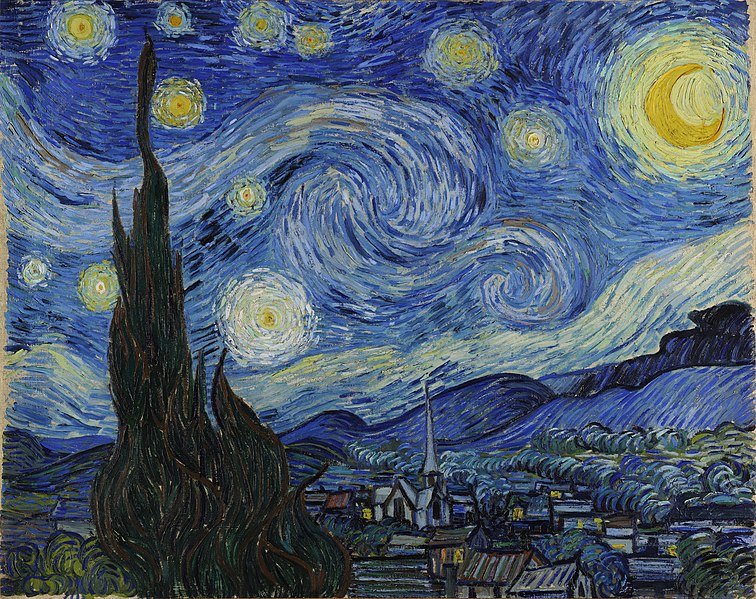
Starry Night, a famous Van Gogh painting some
believe is the result of his hallucinations

While this new study offers great evidence comparing schizophrenia patients with different symptoms, there is still a lot to figure out about this kind of hallucination. Scientists are still working to discover what exact processes lead to cortical thinning and how those processes begin. However, what we do know about auditory verbal hallucinations emphasizes how heavily we rely on our perception of the world around us. We will not ever get to know the thickness of Vincent Van Gogh’s MTG, but the auditory hallucinations Van Gogh experienced were probably the result of his hearing system malfunctioning in some way. Today, many people believe that some of Van Gogh’s most famous decisions and artworks were informed by his hallucinations (Jones, 2016; New York Times Archive, 1981). Modern neuroscience tells us that those hallucinations may have actually been an erroneous interpretation of his own inner dialogue all along. 


References

American Psychiatric Association. (2013). Diagnostic and statistical manual of mental disorders (5th ed.). Arlington, VA: Author.

Binney RJ, Parker GJ, Ralph MAL (2012). Convergent connectivity and graded specialization in the rostral human temporal lobe as revealed by diffusion-weighted imaging probabilistic tractography. Journal of Cognitive Neuroscience 24, 1998–2014.

Catani M, Ffytche DH (2005). The rises and falls of disconnection syndromes. Brain 128, 2224–2239.

Cabeza R, Nyberg L (2000). Imaging cognition II: an empirical review of 275 PET and fMRI studies. Journal of Cognitive Neuroscience 12, 1–47.

Cui Y, Liu B, Song M, Lipnicki D, Li J, Xie S, . . . Jiang T. (2018). Auditory verbal hallucinations are related to cortical thinning in the left middle temporal gyrus of patients with schizophrenia. Psychological Medicine, 48(1): 115-122

Jones, J. (2016). Vincent van Gogh: Myths, madness and a new way of painting. Retrieved from https://www.theguardian.com/artanddesign/2016/aug/05/vincent-van-gogh-myths-madness-and-a-new-way-of-painting

Gogtay N, Giedd JN, Lusk L, Hayashi KM, Greenstein D, Vaituzis AC, Nugent TF, Herman DH, Clasen LS, Toga AW, Rapoport JL, Thompson PM (2004). Dynamic mapping of human cortical development during childhood through early adulthood. Proceedings of the National Academy of Sciences 101, 8174–8179.

Lewis DA, Levitt P (2002). Schizophrenia as a disorder of neurodevelopment. Annual Review of Neuroscience 25: 409–432.

McGuire PK, David AS, Murray RM, Frackowiak RSJ, Frith CD, Wright I, Silbersweig DA (1995) Abnormal monitoring of inner speech: a physiological basis for auditory hallucinations. The Lancet, 346(8975): Pages 596-600,

New York Times Archive (1981) Van Gogh’s Hallucinations. Retrieved from https://www.nytimes.com/1981/07/07/science/science-watch-van-gogh-s-hallucinations.html

Seal ML, Aleman A, McGuire PK (2004). Compelling imagery, unanticipated speech and deceptive memory: neurocognitive models of auditory verbal hallucinations in schizophrenia. Cognitive Neuropsychiatry 9, 43–72.

Shergill SS, Brammer MJ, Williams SCR, Murray RM, McGuire PK (2000). Mapping auditory hallucinations in schizophrenia using functional magnetic resonance imaging. Archives of General Psychiatry 57, 1033–1038

Van Gogh Museum (2016). Shortly before 27 February 1889 In Concordance, lists, bibliography (Documentation). Retrieved from: http://www.vangoghletters.org/vg/documentation.html


Images: 

https://search.creativecommons.org/photos/71b807e7-29fd-445d-95a1-4d282ccf02e5

https://upload.wikimedia.org/wikipedia/commons/thumb/f/f5/Gray726_middle_temporal_gyrus.png/250px-Gray726_middle_temporal_gyrus.png

https://upload.wikimedia.org/wikipedia/commons/thumb/e/ea/Van_Gogh_-_Starry_Night_-_Google_Art_Project.jpg/757px-Van_Gogh_-_Starry_Night_-_Google_Art_Project.jpg

Let’s Van Go(gh) to Arles

Narrow streets, old buildings, and small shops were in sight as I walked with a group of my friends towards the renowned Vincent Van Gogh Café. We were in Arles, a city in the south of France where the Dutch painter Van Gogh lived for more than a year and created some of his best work. Once we reached the square, a yellow café was to our right, and in blue writing “Le Café La Nuit” and “Vincent Van Gogh Café” were spelled out.  Red couches and vases of yellow sunflowers lined the walls. People were rushing in and out, and tourists were taking pictures.

The image of Vincent Van Gogh Café in 2019 compared to Van Gogh’s painting in 1888.

This café, once a place where Van Gogh spent his time painting, now differed from the one depicted in his painting. Chairs were replaced by couches, empty stores fronts were changed to buzzing restaurants and hotels, a few circular tables were swapped for large rectangular tables. Intrigued, we did not stop there. Our next stop was the hospital ward courtyard, where Van Gogh was admitted twice, and created three known paintings.

The courtyard of the hospital ward in Arles (2019), with a replica of Van Gogh’s painting in the foreground.

You might be confused right now if you have not heard about Van Gogh’s story. Hospital ward? Twice? Van Gogh left Paris for Arles because of his mental health. However, after a few months in Arles, his mental health deteriorated. A razor covered with blood in hand, Van Gogh had maimed his ear, after arguing with his house guest (Khoshbin and Katz, 2015). At Hotel Dieu Hospital, Dr. Felix Rey treated Van Gogh, bandaged his ear, and diagnosed him with epilepsy. In a letter Dr. Rey wrote, there were times when Van Gogh “loses his train of thought and speaks nothing but disjointed words… he went to lie down in another patient’s bed and would not leave it… he chased the sister on duty… he went to wash in the coal-box” (Van Gogh Museum). He was then transferred to the asylum Saint-Remy-de-Provence, where Dr. Theophile Peyron, recorded Van Gogh’s medical condition as having “suffered an attack of acute mania with visual and auditory hallucinations that led him to mutilate himself by cutting off his ear” (Van Gogh Museum). So, was Van Gogh certainly epileptic? Or did he suffer from another neurological disorder?

Epilepsy is a neurological condition that is defined by recurrent seizures and can affect people of all ages. Van Gogh was described by doctors as having seizures, which is the reason for the primary diagnosis of epilepsy. However, the best method for diagnosis is through the use of an electroencephalography (EEG), a machine that records the electrical activity of the brain (Guerreiro, 2016). Epileptic patients have unusual activity in their brain cells(neurons), which makes an EEG a good tool to detect epilepsy. However, in 1889, when Van Gogh was diagnosed, the EEG had not yet been discovered. Thus, with no scans to look at, this brings to question whether the diagnosis of Van Gogh was accurate.

EEG image of a normal (seizure-free) brain compared to an EEG image of the brain of an epileptic patient (Ebrahimpour et al. 2012).

Another study revealed that auras are important in diagnosing patients as epileptic (Liu et al., 2017). An aura consists of all the sensations that a patient experiences prior to a seizure. The type of aura the patient experiences conveys what part of the cerebral cortex, outermost region of the brain responsible for decision making and speech, is affected (Liu et al., 2017). Epileptic patients could have sensory (related to the senses) or cognitive auras (related to thoughts), as well as unspeakable feelings (Liu et al., 2017). These characteristics were evident in Van Gogh, since he had auditory and visual hallucinations and he was unable to express his thoughts. However, the findings do not explain the depressive symptoms and the urge to commit suicide that Van Gogh experienced.

The more I look at the symptoms described by the doctors, the more I realize that Van Gogh was more likely a schizophrenic. Schizophrenia is a brain disorder that encompasses hallucinations, delusional thinking and cognitive problems (Seidman and Mirsky, 2017). One study examined the effects of depression and cognitive impairment on adults with schizophrenia (Raykeer et al., 2019). Patients who had schizophrenia had increased depression and cognitive impairments, which they measured through “quality of life exams,” a common well-known method. Both depression and cognitive impairments were observed in Van Gogh, according to the medical records written by Dr. Peyron. Further, individuals with schizophrenia lack empathy, are unable to understand what other people are feeling based on gestures, and have poor problem-solving skills (Couture et al., 2006). All of these symptoms were manifested by Van Gogh. Therefore, it is likely that he may have been schizophrenic, although there is no conclusive evidence to determine his neurological condition.

Now, as I continue walking towards the river in Arles, I see a replica painting of Van Gogh’s “Starry Night, 1889,” outside a gallery. This was a painting he made through his window when he was institutionalized at Saint Remy Asylum. Some people say that it was a visual hallucination because Van Gogh’s room did not have a view of the city and the trees were not shaped like flames, nor did the stars whirl as they appear in his painting. So, what was Van Gogh’s medical condition? The question remains unanswered, but if you asked me, I would say all signs point to schizophrenia.

Image of Van Gogh’s Starry Night 1889 painting.


References:

Couture, S. M., Penn, D. L., & Roberts, D. L. (2006). The functional significance of social cognition in schizophrenia: a review. Schizophrenia bulletin32(suppl_1), S44-S63.

Ebrahimpour, R., Babakhan, K., Arani, S. A. A. A., & Masoudnia, S. (2012). Epileptic seizure detection using a neural network ensemble method and wavelet transform. Neural Network World22(3), 291.

Guerreiro, C. (2016). Epilepsy: Is there hope? Indian Journal Of Medical Research144(5), 657.

Khoshbin, S., & Katz, J. (2015). Van Gogh’s Physician. Open Forum Infectious Diseases2(3), ofv088.

Liu, Y., Guo, X. M., Wu, X., Li, P., & Wang, W. W. (2017). Clinical Analysis of Partial Epilepsy with Auras. Chinese medical journal130(3), 318.

Pascal de Raykeer R, e. (2019). Effects of depression and cognitive impairment on quality of life in older adults with schizophrenia spectrum disorder: Results from a multicenter study. J Affect Disord. 256, 164-175.

 Seidman, L. J., & Mirsky, A. F. (2017). Evolving notions of schizophrenia as a developmental neurocognitive disorder. Journal of the International Neuropsychological Society23(9-10), 881-892.

Van Gogh Museum. (2009, October). Vincent Van Gogh The Letters. Van Gogh Museum. Retrieved from http://vangoghletters.org/vg/letters.html


Paintings from:

Van Gogh V. (1889). The Starry Night. Retrieved from

https://www.vincentvangogh.org/starry-night.jsp

Van Gogh V. (1888). Café at Night. Retrieved from

https://www.vincentvangogh.org/cafe-at-night.jsp


Lust for Answers

This past weekend, our group went to Provence, a province in southeast France, and visited the city of Arles where Vincent van Gogh lived for two years painting some of his most famous works such as Yellow House, Starry Night Over the Rhone, and Bedroom in Arles.

A map of some of the locations in Arles where van Gogh painted some of his most famous works.

Before going there, we saw in class the movie, Lust for Life, a 1950’s biographical movie about Vincent van Gogh’s life highlighting his interactions with other painters, his family, and his surroundings (Lust for Life – Trailer, n.d.). The movie touches on Van Gogh’s lifelong mental strife showing that while we revere him as an artistic genius now, very few people understood him including himself.

It seemed the depression that Van Gogh experienced subsided according to his letters to his family and friends, but in the movie, they show the manic way he painted constantly covered in paint and obsessed with catching the light to paint landscapes and field laborers. When the fall and winter came around, he could not go outside expressing how he felt trapped. His condition worsened where outside painting did not work anymore leading up to him to cut off his ear with a variety of possible reasons that no one could confirm. He eventually was admitted to a hospital where his hallucinations continued with blocks of time missing from his memory and his alcohol abuse addressed. He still continued to paint famous pieces such as The Courtyard of the Hospital at Arles 1889 that are preserved to this day.

A picture at the hospital courtyard where van Gogh was attempted to after cutting off his ear.

We looked at his doctor’s notes categorizing his condition as epilepsy because of his ongoing non-lucid episodes, so we started looking into different mental conditions that related back to the ones we know today as major depression disorder, bipolar disorder, schizophrenia, and more. This eventually led us to see what type of treatments would be available for the people with dementia praecox: a term coined by Emil Kraepelin to describe lesions in the cerebral cortex that mild dementia (Adityanjee et al., 1999). I couldn’t find much in terms of treatment, but it got me thinking about what we have today to help alleviate the effects of mental illnesses such as bipolar disorder, schizophrenia, and depression. As well as my interest being piqued through exploring Van Gogh’s life, there is a high probability I will see these novel practices implemented in the future.

The School of Nursing at Emory does a good job of teaching us the evidence-based practices that we follow for patient safety and comfort, but the patients have the autonomy in most cases to deny treatment, do something different than recommended to treat their ailments , or ask about new upcoming treatments. Because of this, it is important to know recent research about various types of treatment practices to be better support the patients.

van Gogh’s self portrait fading away during the Atelier des Lumieres of all of his works.

One that was really interesting to find out was the possible implementation of brain-derived neurotrophic factor (BDNF) to help treat neurodegenerative diseases as well as mental health disorders. It is a growth factor that is used in neurogenesis or the formation of new neurons which is not common for adults; in most of the brain, there are no new neurons created from the ones at birth, but there are some brain areas that still have new neurons created which is where growth factors like BDNF are used those new neurons (Bathina and Das, 2015). This is also used for synaptic plasticity in which there is a consistently strong or diminished communication between the neurons depending on how strength and importance of the signal is between the two neurons (“What Is Synaptic Plasticity?”). There is also evidence that a depleted amount of the class of factors BNDF belongs to can possibly be an indicator for neurological disorders such as Parkinson’s disorder and Alzheimer’s (Bathina and Das, 2015). While BNDF has the ability for synaptic plasticity, a study done with mice found that inhibition of one of the receptors BNDF can bind to shows a decrease in long term depressive behaviors without affecting its synaptic plasticity function in other brain areas (Woo et al., 2005). Researchers also theorize that people with reduced BDNF levels might have a decreased synaptic plasticity in the hippocampus which prevents the body from going back to homeostasis taking them out of their stress related depressive states (Phillips, 2017). The second type of receptors that BDNF does the opposite effect by producing synaptic plasticity; this receptors’ activation and an BDNF increase is seen in the presence of certain antidepressive pharmacologic therapies (Phillips, 2017).This is now being used as an indicator for future drug therapies as a measure of effectiveness.

A watercolor painting I did in class depicting the sensory neurons in the eye.

Going away from the pharmacological side, I started to think about Van Gogh and how his art was a source of peace and strife for him. At some point, painting couldn’t help in him in the way it did before. This is not to discredit the effects that art and other alternative therapies have on supporting those with symptoms similar to his; a study had 58 patients diagnosed with schizophrenia do art therapy twice a week for twelve weeks (Montag et al., 2014). They found that those who had committed to the program had less negative symptoms which include a loss of interest and a lower affect as well as less positive symptoms of schizophrenia such as auditory hallucinations compared to the control group who did not receive the art therapy (Montag et al., 2014)  (“Symptoms,” 2017). This support the idea that Van Gogh’s art was a therapeutic event for him up until everything became too much. It’s fascinating to how we reverie Van Gogh’s coping mechanism after his death with his few family and friends supporting his ability to paint. It makes you think about those that we have forgotten about who are tucked away in our society creating the next artistic masterpiece of our time.


References

Adityanjee, Aderibigbe, Y. A., Theodoridis, D., & Vieweg, W. V. R. (1999). Dementia praecox to schizophrenia: The first 100 years. Psychiatry and Clinical Neurosciences, 53(4), 437–448. https://doi.org/10.1046/j.1440-1819.1999.00584.x

Bathina, S., & Das, U. N. (2015). Brain-derived neurotrophic factor and its clinical implications. Archives of Medical Science: AMS, 11(6), 1164–1178. https://doi.org/10.5114/aoms.2015.56342

Lust for Life – Trailer. (n.d.). Lust for Life – Trailer. Retrieved from https://www.youtube.com/watch?v=WUHL0h_kQ6s

Montag, C., Haase, L., Seidel, D., Bayerl, M., Gallinat, J., Herrmann, U., & Dannecker, K. (2014). A Pilot RCT of Psychodynamic Group Art Therapy for Patients in Acute Psychotic Episodes: Feasibility, Impact on Symptoms and Mentalising Capacity. PLoS ONE, 9(11). https://doi.org/10.1371/journal.pone.0112348

Phillips, C. (2017). Brain-Derived Neurotrophic Factor, Depression, and Physical Activity: Making the Neuroplastic Connection. Neural Plasticity, 2017.https://doi.org/10.1155/2017/7260130

Symptoms. (2017, October 23). Retrieved June 10, 2019, from nhs.uk website: https://www.nhs.uk/conditions/schizophrenia/symptoms/

What is synaptic plasticity? (2016, November 22). Retrieved June 9, 2019, from https://qbi.uq.edu.au/brain-basics/brain/brain-physiology/what-synaptic-plasticity

Woo, N. H., Teng, H. K., Siao, C.-J., Chiaruttini, C., Pang, P. T., Milner, T. A., … Lu, B. (2005). Activation of p75NTR by proBDNF facilitates hippocampal long-term depression. Nature Neuroscience, 8(8), 1069–1077. https://doi.org/10.1038/nn1510

Picture #1: [Screenshot of the walking tour of Van Gogh’s art in Arles]. Retrieved from https://www.google.com/maps/d/u/0/viewer?mid=1014-AkOjbBzXEQQLcxz8NGxa10Oo1bGN

Picture #2 and #3: Taken by me

Picture #4: Painted and picture taken by me