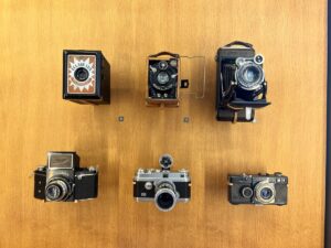
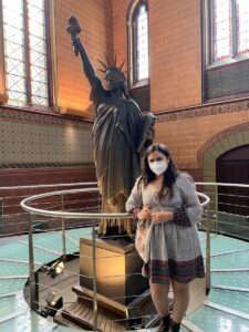
I have always loved taking photos. When preparing for any trip, the first things in my suitcase are always my cameras! My goal for Paris was to be able to document all the beautiful buildings, delicious food, and fun experiences through my camera lens. A few days ago when I first heard about our trip to the Musée des Arts et Métiers, or Museum of Arts and Crafts, on Wednesday, June 8th, I wasn’t sure what to expect. However, I was pleasantly surprised to find rows and rows of scientific instruments and inventions in front of me. The contraptions ranged from communication devices to modes of transportation and even architectural models. My favorite room as a whole was the one that housed the French replica of the Statue of Liberty, airplane models, and antique cars at the end of the museum (Figure 1). But if I had to choose one singular exhibit that called my attention the most, it was the showcase of all the different cameras throughout the years (Figure 2).

It is fascinating to think about the major technological improvements that have been made over the past 100 years and how the invention of one item can pave an avenue of inspiration for so many other products. The development of the camera not only served as a tool for leisure and documentation but also allowed improvements in the medical field. Today, cameras are used in many procedures including neuroendoscopies. A neuroendoscopy was not a technique I was very familiar with, but it is a minimally invasive surgery tool that allows for tissue sampling in the brain usually because of brain tumors. In a retrospective descriptive study conducted by Deopujari, Shroff, Karmarkar, & Mohanty in 2022, 27 previous procedures were analyzed that utilized either endoscopic tumor biopsy (ETB) or endoscopic third ventriculostomy (ETV) techniques to treat pediatric patients. Children were laid in an upright position with their heads turned to a 20-degree angle to the left such that the right side was exposed for these procedures. Researchers were able to insert the endoscope and perform the method with just a 4-6 cm coronal suture. The study showed that the accuracy of these procedures in children with pineal region tumors “has been above 75%”, which is a statistically significant value. Neuroendoscopic biopsy as a technique is becoming much more common in the medical field as it has proven itself as a safe and effective procedure with minimal invasiveness. I can expect this technology to expand even further in the next few years as it becomes more and more effective.

References:
Deopujari, C., Shroff, K., Karmarkar, V., & Mohanty, C. (2022). Neuroendoscopy in the management of pineal region tumours in children. Child’s Nervous System. https://doi.org/10.1007/s00381-022-05561-0
