A Snapshot into Medical Technology

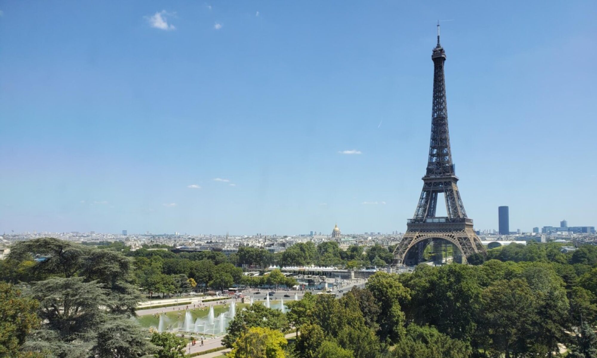
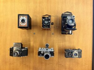
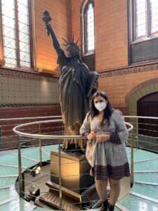
I have always loved taking photos. When preparing for any trip, the first things in my suitcase are always my cameras! My goal for Paris was to be able to document all the beautiful buildings, delicious food, and fun experiences through my camera lens. A few days ago when I first heard about our trip to the Musée des Arts et Métiers, or Museum of Arts and Crafts, on Wednesday, June 8th, I wasn’t sure what to expect. However, I was pleasantly surprised to find rows and rows of scientific instruments and inventions in front of me. The contraptions ranged from communication devices to modes of transportation and even architectural models. My favorite room as a whole was the one that housed the French replica of the Statue of Liberty, airplane models, and antique cars at the end of the museum (Figure 1). But if I had to choose one singular exhibit that called my attention the most, it was the showcase of all the different cameras throughout the years (Figure 2).

Figure 1. Here you can see the advancements of the digital camera over time.


It is fascinating to think about the major technological improvements that have been made over the past 100 years and how the invention of one item can pave an avenue of inspiration for so many other products. The development of the camera not only served as a tool for leisure and documentation but also allowed improvements in the medical field. Today, cameras are used in many procedures including neuroendoscopies. A neuroendoscopy was not a technique I was very familiar with, but it is a minimally invasive surgery tool that allows for tissue sampling in the brain usually because of brain tumors. In a retrospective descriptive study conducted by Deopujari, Shroff, Karmarkar, & Mohanty in 2022, 27 previous procedures were analyzed that utilized either endoscopic tumor biopsy (ETB) or endoscopic third ventriculostomy (ETV) techniques to treat pediatric patients. Children were laid in an upright position with their heads turned to a 20-degree angle to the left such that the right side was exposed for these procedures. Researchers were able to insert the endoscope and perform the method with just a 4-6 cm coronal suture. The study showed that the accuracy of these procedures in children with pineal region tumors “has been above 75%”, which is a statistically significant value. Neuroendoscopic biopsy as a technique is becoming much more common in the medical field as it has proven itself as a safe and effective procedure with minimal invasiveness. I can expect this technology to expand even further in the next few years as it becomes more and more effective.

Figure 2. Me posing by a French replica of the Statue of Liberty!


References: 

Deopujari, C., Shroff, K., Karmarkar, V., & Mohanty, C. (2022). Neuroendoscopy in the management of pineal region tumours in children. Child’s Nervous System. https://doi.org/10.1007/s00381-022-05561-0

Surprise ! It’s a soccer match!

On Friday, June 3rd, my group presented article 3 which focused on the association between participation in professional rugby and mild traumatic brain injuries. Our group introduced the article with a brief presentation on how the game of rugby works, including the different player positions and responsibilities on the field. Leaving class as rugby experts, we were excited to travel to the Stade de France to watch a rugby match between France and Denmark. 

As my roommate and I were walking toward the stadium from the metro, we realized the crazy number of fans that were waiting in line to get in. After getting the flag of France painted on our cheeks to prove team pride and blend in, we made our way into the enormous stadium to search for our seats. Minutes before the game started, we finally realized that the field setup did not match the rugby setup. Instead of the H-shaped goal posts, there were two soccer goals. 

As I took this photo, the realization hit that this looked suspiciously like a soccer game.


Although it wasn’t what we were expecting, it was certainly a nice surprise! We were originally tasked to pick out a rugby player and keep track of each time they suffered a hit to the head for the duration of the match. Shifting our task to the soccer match, we noticed that there were few to no instances that the players received a hit to the head unintentionally. Instead, the players seemed to intentionally hit the soccer ball with their heads as part of a play. The combination of so much movement across the field and no real-time replays resulted in an estimation that the French team’s individuals had this type of hit at least 7 times. 

Jewel, Khushi, Solanch, and me when we found out we were at a soccer match!


Following the preliminary discussion of mTBI in professional rugby players and reflecting on the soccer match, I was curious to learn more about the prevalence of concussions. According to Bunc et al.,2017, a professional soccer player hits the ball with their head between 6-17 times per match. As with professional rugby players, a team of researchers also found “increased evidence for brain atrophy”, or loss of neurons and neuronal connections when they compared imaging to a non-soccer player control group (Tysvaer et al., 1989). These specific findings led to recommendations on emphasizing head injury assessment and treatment, using proper head play techniques, and using a different size ball (3-5) depending on the age ranges of the players. It’s interesting to learn and see how neuroscience findings translate to real-world implications.

Bunc, G., Ravnik, J., & Velnar, T. (2017). May Heading in Soccer Result in Traumatic Brain Injury? A Review of Literature. Medical archives (Sarajevo, Bosnia and Herzegovina), 71(5), 356–359. https://doi.org/10.5455/medarh.2017.71.356-359 

Tysvaer, A. T., Storli, O. V., & Bachen, N. I. (1989). Soccer injuries to the brain. A neurologic and electroencephalographic study of former players. Acta neurologica Scandinavica, 80(2), 151–156. https://doi.org/10.1111/j.1600-0404.1989.tb03858.x 


A Visit With Unique Wax Models

Today we ventured to the Musée de Moulages for the second time. Although it was an optional excursion, I was excited to go and see the different skin disease and conditions. Traveling to the museum allowed us to walk through a new arrondissement of Paris and see a beautiful canal. Held within hospital grounds, we saw patients enjoying the outdoors and doctors taking a break or walking to a neighboring building in the hospital.

One of the photographs displayed in the entryway of the museum to illustrate a dermatological condition.

Upon entering the museum, we saw drawings and paintings of patients with different skin conditions. In the entryway, there was a sculpture of Louis IX, the ancestor of Henry IV. Upstairs housed the real exhibit: over 4,800 castings of all types of dermatological problems, from syphilitic chancres to eczema to elephantiasis. Jules Baretta is the one who first started making the wax dermatological models after his great success with his realistic models of fruit. Despite these fascinating displays, we were strictly prohibited from taking photos, as they came from moldings of real patients, so the museum is protecting their privacy. I think it is important that the museum values the privacy of the patients so strongly, even though the patients are most likely dead.

A selfie with the different photographs and drawings of dermatological patients.

I was most fascinated by the syphilis exhibition, one of their largest displays with 442 wax models. In class, we learned about neurosyphilis and ocular syphilis and their corresponding symptoms. As I was a part of the group who presented on this target article, I had the pleasure of finding Google Images of these symptoms, such as the chancres. I realized that Google Images only displayed the mild versions of the symptoms. The extent to which syphilis can affect the body is truly horrifying, most of which I don’t think I’ll be able to unsee. I think modern medicine has come a long way since the creation of some of these castings. Doctors can detect syphilis sooner and treat it easily with penicillin, so the symptoms are not as severe.

It is important that these skin diseases are still understood and studied, even though we use photographs instead of wax models, because there has been a rise of syphilis in the past two decades, specifically in women and congenital syphilis. The museum displayed the presence of congenital syphilis in babies, and it was very unfortunate. Testing for neurosyphilis can still be difficult, but doctors normally rely on the CSF Venereal Disease Research Laboratory with a higher titer cutoff to diagnose neurosyphilis (Chow, 2021). Without modern medicine and antibiotics, a lot more people would die from syphilis and neurosyphilis.

Chow F. (2021). Neurosyphilis. Continuum (Minneapolis, Minn.)27(4), 1018–1039. https://doi.org/10.1212/CON.0000000000000982

Monet On My Mind

Last Sunday on June 12th I went to Giverny, Paris to visit the gardens and house of Claude Monet. My friend, Jewel and I took the SNCF train from Saint Lazare to Vernon-Giverny station from which we rented a bicycle from a nearby shop to go to Giverny. We set sail on our bikes along the French countryside and rode our way to Monet’s house.

We reached the most beautiful little town I have ever seen! The whole town was filled with colorful flowers ranging from Lillies to peonies to even white roses. We got our tickets and went straight to Monet’s gardens. The garden’s entrance felt like being in an enchanted maze, filled with the sound of birds and a feel of fresh air. I felt rejuvenated walking through Monet’s gardens and the town of Giverny itself, and I wondered why I felt so calm and energized simultaneously. We talked about neurotechnology in class- feelzing- a company manufacturing a device that stimulates the brain to feel calm and energized at the same time, and I wondered how I felt this way without a device. The only possible reason I could think of was Nature and its effects on the mind.

Riding along the Seine to get to Giverny. We rode for 6 km along the river and hillside until we reached the small town of Giverny.

It is common knowledge that being in nature helps reduce anxiety and stay calm- a sort of very natural meditation many of us experience. It is also commonly perceived that people who tend to live near a lot of nature including trees, forests, lakes, sea, mountains, etc., or interact with nature daily are happier than those who do not interact with or in nature. According to White et al. (2013), increasing green space in urban areas promotes reduced mental distress and higher well-being on an individual level. There exists an interesting theory known as the Biophilia hypothesis, proposed by Harvard biologist Edward O Wilson, that suggests that humans have an innate affiliation to other living organisms, and this spans across all cultures and countries (Selhub, Logan 2012). Biophilia is a hypothesis that may be able to explain the connection many of us feel when we are amidst nature.

According to Cox et al. (2017), increased vegetation cover and bird sound abundance were positively associated with a lower prevalence of depression, anxiety, and stress. Biologically, this increase in mental health is due to the various smells that we experience in nature including flowers, nectar, water, and also the sounds we experience from various birds, insects, animals, and moving water which helps us recover from stress than sounds of urban life, (Alvarsson et al., 2010).

Me enjoying the little pond and forest behind Monet’s house which influenced a lot of his paintings. One of his most famous series of paintings- The water Lilly series was inspired by the water lilies and bridge behind his house.

Overall, this was one of the best trips I have experienced until now and I would recommend going to Giverny to anyone looking for a “secret garden” escape from their busy life. Cycling along the Seine across the country hillside was a great therapeutic choice after a hectic week of work and papers.

References:

https://www.ncbi.nlm.nih.gov/pmc/articles/PMC2872309/

https://books.google.fr/books?hl=en&lr=&id=CFd_7ujLlwgC&oi=fnd&pg=PA1&dq=being+in+nature+and+the+brain&ots=I848kBeiEQ&sig=EY9pICdBZlVPamw6uE0Gp7erWu0&redir_esc=y#v=onepage&q=being%20in%20nature%20and%20the%20brain&f=false

https://journals.sagepub.com/doi/abs/10.1177/0956797612464659

https://academic.oup.com/bioscience/article/67/2/147/2900179?login=true

https://jech.bmj.com/content/72/10/958.abstract

A COVID-19 Memoir: “Prison” with a view

This week I spent most of my time in Hell! As many of you know, I contracted COVID-19 last week and had to quarantine for 5 days. My experience was awful. There is no sugar coating it. The virus drained me both physically and mentally.

Prior to quarantining, we went to the Soccer game which was amazing but I’m pretty sure that that’s the day of first exposure resulting in me contracting Covid.

Here is me in a state of blissful ignorance prior to getting my positive result.

Fast forward to this past Monday night when I started to feel low energy. I knew that my immune system was on alert but I thought it was just a sinus problem. The next day, however, I started to feel some serious fatigued and was told to get tested. When I got the positive result, I was heartbroken. This was my first time contracting COVID so I didn’t know what to expect.

The physical symptoms were hard but manageable but the mental ones really hit me. During that time, I had felt isolated, depressed, and lonely but also simultaneously stressed about missing all this work. In thinking about this loneliness I searched for any additional information. From what I found I learned that the “lonely areas” in the brain are the amygdala as it is the emotion-center of the brain but also the Nucleus Accumbens which provides a positive reward aspect during social feedback (Lieberz et al. 2021). I also learned the NAcc is less activated during times of loneliness which may contribute to an overall lack of motivation( Lieberz et al. 2021). With no social feedback and the same environmental stimulus, no wonder why I lacked the motivation to do anything. I think it was the loneliness that was preventing me from working


One thing that helped me through all this loneliness was all the people supporting me I’m extremely grateful for my classmates who reached out and checked in on me and sent wishes to get better. These very kind messages did wonders for my immune system as well as my overall hope of getting through this. I’m even more grateful to my roommate Adway for making me soups for the past couple of days and Duke for disinfecting any surface that I touched to ensure that I would spread this horrid virus to anyone else. I’m also really grateful for my Mom and partner who were rooting for me through this dark time. Also, shout out to the two Squishamallows that my partner packed for me. These guys are the real pillars of my mental health!!

Here are my support fruits that got me through it! Left is Maui and right is Ximena. Also, the cool view where I spent most of my time people-watching can be seen in the background.

Lieberz, J., Shamay-Tsoory, S. G., Saporta, N., Kanterman, A., Gorni, J., Esser, T., Kuskova, E., Schultz, J., Hurlemann, R., & Scheele, D. (2021). Behavioral and neural dissociation of social anxiety and loneliness. https://doi.org/10.1101/2021.08.25.21262544 

Rugby (and European Football) and CTE

by Samantha Feingold

“Does anyone know what Rugby actually is?” This question floated around in several variations the week before we were to attend a France vs. Denmark rugby match.

“What?” asked our waiter several times before it was evident we weren’t pronouncing it well enough for him to understand. Lauren gestured throwing a rugby and he finally understood, but said he didn’t know the sport well enough to explain. While the restaurant is nearby our apartment and we have eaten there enough times for this waiter to recognize us when we walk past, an Italian restaurant maybe wasn’t the best place to ask about rugby.

“It’s like football but without helmets,” said a peer. It is important I emphasize at this time she was referring to American football. This statement left me more confused as helmets and padding seem to be the most important part of football, a high contact sport. Research on retired football players with chronic traumatic encephalopathy (CTE) has amplified in conjunction with the increase of media coverage on cases of severe cognitive deterioration and changes in mood in famous players (Buckland et al., 2019). However, there are minimal studies on CTE and long-term negative effects on the brain for rugby players (Buckland et al., 2019).

Prior to the match, my peers presented a crash-course on rugby and an in-depth journal article review on rugby and CTE (Zimmerman et al., 2021). It quickly became evident that rugby is quite intense, and it was likely there would be many head collisions.

When we finally arrived at Stade de France, all the fans were bouncing with excitement and kids ran around with French-flag face paint. It wasn’t until we settled in our seats and the pre-match rituals concluded that we realized the peculiar goals and that what in front of us resembled European football far more than having any parallels to American football (Figure 1).

Figure 1. The European football field at Stade de France for the Denmark vs. France match.

While we did not see rugby that evening, we were lucky enough to watch a qualifying match for the World Cup (Figure 2)! Within minutes of realizing this was soccer and not rugby, it became evident that instead of being tackled, there was risk for brain injury from repeatedly heading the soccer ball. The potential force exerted from heading ranges from 500-1200 Newtons (Teymouri et al., 2012) and is equivalent to a maximum of over 250 pounds-force (lbf). While this is not quite the impact of a moving 300+ pound American football player, the European football players are (like the elite rugby players) not wearing helmets. Thus, while soccer is very entertaining, it is important to properly teach young players how to head a soccer ball and be aware of the possible injury.


Figure 2. Samantha Feingold, Lauren Cobitz, and Dr. Easterling photographed at the Denmark vs. France soccer match.

Works Cited

Buckland, M.E., Sy, J., Szentmariay, I. (2019). Chronic traumatic encephalopathy in two former Australian National Rugby League players. acta neuropathol commun 7, 97. https://doi.org/10.1186/s40478-019-0751-1

Teymouri, M., Sadeghi, H., Nabaei, A., & Kasaeian, A. (2012). The relationship between
biomechanical-anthropometrical parameters and the force exerted on the head when
heading free kicks in soccer. Archives of trauma research, 1(1), 44–48. https://doi.org/10.5812/atr.5307

Zimmerman, K. A., Laverse, E., Samra, R., Yanez Lopez, M., Jolly, A. E., Bourke, N. J.,
Graham, N., Patel, M. C., Hardy, J., Kemp, S., Morris, H. R., & Sharp, D. J. (2021).
White matter abnormalities in active elite adult rugby players. Brain
communications, 3(3), fcab133. https://doi.org/10.1093/braincomms/fcab133

Christianity, COVID, and Cathedrals

On June 11th, 2022, Sam, Rachel, Cynthia, and I visited Reims, France with Aman, one of my friends from Emory. During our visit, we saw the Notre-Dame Cathedral of Reims, which translates to “Our Lady of Reims.” This beautiful Roman Catholic cathedral, dedicated to the Virgin Mary, was the traditional location for the coronation of the kings of France. The cathedral was founded in the 5th century and has gone through numerous restorations throughout the past few centuries, including after being damaged in the French Revolution and in World War I, when it was struck by more than three hundred artillery shells. I was not only awed by beautiful building but also the deep-rooted history of Christianity in France. Christianity is as much embedded France’s history as it is ingrained in the culture and everyday lives of French citizens. For example, I found it interesting to learn that the grocery store near our apartment does not sell alcohol on Sundays.

Figure 1. A photo of Sam, Cynthia, Rachel, and I in front of the Notre Dame Cathedral of Reims

While visiting the cathedral, I reflected on the articles we discussed in NBB 402W about COVID-19, which sparked my interest in learning about the connection between religion and the pandemic since religious services quickly became hotspots for the virus. I wondered whether the illness made people feel more connected to their religious practices, so I did some research regarding the pandemic and religion. One Polish study found that most citizens surveyed declared faith/spiritually was important to them, but their faith did not strengthen in the face of the pandemic. They found that a higher percentage of Catholic respondents believed in the protective power of faith compared to other denominations and more Catholic respondents also declared that faith had a crucial role in their life (Kowalczyk et al., 2020). Despite this study showing that the pandemic did not increase spiritual belief, religion is obviously an important part of the European lifestyle. Studies have shown neurobiological changes correlated with religion and spirituality specifically in medial frontal cortex, orbitofrontal cortex, precuneus, posterior cingulate cortex, default mode network, and caudate (Rim et al., 2019), which could explain the importance of religion in people’s lives.

Figure 2. Picture of the stain glass windows on the inside of the cathedral

Although the outcomes of the pandemic study were not what I was expecting, I found it interesting to learn more about Christianity and Catholicism in Europe, as well as the brain changes association with religion. Overall, this visit allowed me to feel more connected to my late grandparents on my dad’s side, who were raised Catholic.

Citations:

Kowalczyk O, Roszkowski K, Montane X, Pawliszak W, Tylkowski B, Bajek A. Religion and Faith Perception in a Pandemic of COVID-19. J Relig Health. 2020;59(6):2671-2677. doi:10.1007/s10943-020-01088-3

Rim, J. I., Ojeda, J. C., Svob, C., Kayser, J., Drews, E., Kim, Y., Tenke, C. E., Skipper, J., & Weissman, M. M. (2019). Current Understanding of Religion, Spirituality, and Their Neurobiological Correlates. Harvard review of psychiatry27(5), 303–316. https://doi.org/10.1097/HRP.0000000000000232

Eiffel Tower – Work of Art or Eyesore?

On Wednesday, June 1st, a group of students took advantage of having no major assignments due the next day and spent the evening at the Eiffel Tower. All walking towards the tower from different directions due to the locations of our various metro stops, we met under the center of the tower which we quickly learned was not the ideal location. We relocated to the lawn where we found an open area of grass to have our picnic. I enjoyed my caramel crepe while others were feasting on bread and cheese. Little did we know that we had the best photographer among us (Jacob) who gave us a little spontaneous photoshoot gifting us the ability to remember all the fun we had that night.

Photo by Jacob of me with Cynthia, Sam, and Lauren in front of the Eiffel Tower.

On our first day in Paris we did a boat tour as a class and when we passed the Eiffel tower our guide shared some interesting history about it. The tower’s construction was complete in 1889 in time for the World’s Fair and was not meant to be a permanent structure. When it was first built, Parisians were quite critical of the tower claiming that it was an eyesore and did not fit in with the city. However, despite this opinion of the locals, the Eiffel Tower has become one of the most famous landmarks and is the staple of the city.

This got me thinking about the neuroscience behind the perception of beauty. In previous classes, I have learned about perception of beauty in faces/people and the brain regions involved in that but did not know much about how this applied to objects such as buildings or art. In searching the literature, I was introduced to the field of neuroaesthetics (Iigaya et al., 2020). Neuroaesthetics explores the neural basis for judging art, of which architecture and monuments fall under. Early studies found that the brain regions involved in viewing pleasant aesthetics were also involved in reward pathways. This suggests that looking at an aesthetically pleasing work of art elicits a similar response to other rewarding stimuli. They also explained that assigning aesthetic value is done by breaking a stimulus down into elementary features to be processed. This is a concept I am familiar with from NBB 301 when we discussed sensory processing.

Eiffel Tower lit up as the sun is setting.

In my very unprofessional opinion, I thought the Eiffel Tower was pretty when it was lit up, but during the day it was more exciting due to its significance rather than its beauty.

Reference:

Iigaya, K., O’Doherty, J. P., & Starr, G. G. (2020). Progress and Promise in Neuroaesthetics. Neuron, 108(4), 594–596. https://doi.org/10.1016/j.neuron.2020.10.022

A Tough Loss (on all accounts)

By Ally Grubman

Last Friday the class took a trip to a French Rugby match, or so we thought. About 2 minutes into the game, there were comments flying around our NBB section like “Are we sure this is rugby?”, “When are they going to pick up the ball??”, and my personal favorite, “Guys, they’re just warming up with soccer, don’t worry.” Yep, you guessed it, we made our way to the France vs. Denmark fútbol (soccer) game. So awkward for us.

At the beginning of the soccer game, before the big revelation.

But even though we went thinking it was a rugby game, there was still some relevance to our class that gave me further insight into what we’ve been learning. In our NBB402W class last week, we read a paper on rugby players and the correlation between concussions and later cognition and neurodegenerative illnesses. Although we didn’t get to actually see any rugby players get head injuries, we did however see lots of soccer players use their heads to hit the ball. While this might not be as aggressive or harmful as rugby, there’s no way it’s good for you. For instance, Broglio et al. (2003) found that over a 300-game career, a soccer player experiences roughly 2000 headers, which can add up to cause a lot of damage. This only includes games, not even considering those sustained during practice. This repeated injury can lead to real problems in the future, specifically neurodegeneration. When I say neurodegeneration and neurodegenerative diseases, think: Alzheimer’s, ALS, and Parkinson’s (and so many more). This is something we also found in our rugby paper. Rugby players are rarely required to wear protective or effective helmets. Additionally, since it is very much a contact sport, there has been an increased need for research on the implications of playing such a dangerous game. Tomasin et al. (1989) outline the importance of more awareness on the topic, specifically concerning rugby players and the risk they are putting themselves in. Coaches, physicians, and players need to be more aware of the dangers and the serious possibility of brain damage and degeneration in the player’s future.

A group selfie at the beginning of the game to commemorate the fun time we all had!

However, while we counted the headers and enjoyed the game, we had a great time hanging out in a non-school setting, eating some very overpriced stadium food, and laughing about the fact that we were, in fact, not at a rugby game. It’s too bad that France lost, but all in all, it was a great game and a ton of fun for all of us!!

Citations:

Broglio, S. P., Ju, Y. Y., Broglio, M. D., & Sell, T. C. (2003). The Efficacy of Soccer Headgear. Journal of athletic training, 38(3), 220–224. 

Tomasin, J. D., Martin, D. F., & Curl, W. W. (1989). Recognition and Prevention of Rugby Injuries. The Physician and sportsmedicine, 17(6), 114–126. https://doi.org/10.1080/00913847.1989.11709809

Dermatology includes Cyclops

Last week, the class attempted to visit the Musee des Moulages, but we were turned away as a result of a scheduling error. Our tickets were for the “7th” of June rather than the “1st.” Today, we bravely made the trek back to see the unique collection of wax dermatological models.

The Hospital Saint-Louis was ordered to be built by Henri IV who wanted plague victims to be treated outside the walls of the capital. I was able to get a selfie with him 🙂

Henri IV and I almost touch hands. Historical moment.

This same hospital eventually hosted the genius of Jean-Louis Alibert who introduced the new medical discipline of dermatology. The idea to represent pathologies in three-dimensional representations was inspired by Jules Baretta who made models of fruit from paper board. According to the museum guide, Baretta made his first wax dermatological model in 1867, and eventually, the museum of wax models was established as a learning tool for the students of the hospitals. 

Apparently, the Hospital Saint-Louis models were the inspiration for similar museums across the world and established a reputation that manifested in over 4,800 pieces today–the largest collection of its kind in the world. It is interesting to think that the majority of visualizations that students have access to today are primarily digital, and it may even be the case that the future will allow augmented reality renditions of these models.

Walking around the exhibit, there were many genital representations marred by a variety of disfigurations. I had a difficult time discerning what the diseases were (the descriptions were in French and there was no tour or explanation), but I definitely saw multiple sections dedicated to syphilis. This closely relates to our class discussion of syphilis wherein the disease presents itself in the skin as well as the forms of neuro and optical forms distinguished in our paper. 

“The first symptom of primary syphilis is a usually painless open sore called a chancre (pronounced “shanker”). The chancre can appear within 10 days to 3 months (usually 2 to 6 weeks) after exposure.” As such, there were plenty of wax models of chancres throughout the exhibit, but I was not allowed to take any pictures as the majority of them were on the first floor by the supervisors.

However, on the second floor, I had a little more liberty to take a picture for the benefit of the class–a notable exhibit I saw was one dedicated to “abnormalities,” and I saw a model of a human “cyclops” baby. Again, the museum had a no photography policy, but I took one for the class, for science. 

[Warning, graphic image below]

This is a graphic model of cyclopia–more information is available in the link below.

Here is what I found in research about cyclopia:

“Cyclopia (also cyclocephaly or synophthalmia) is a rare form of holoprosencephaly and is a congenital disorder (birth defect) characterized by the failure of the embryonic prosencephalon to properly divide the orbits of the eye into two cavities. It is the severest facial expression of the holoprosencephaly syndrome.1 Its incidence is 1 in 100 000 in newborns.”

This condition is interesting to read through especially considering our discussion of ‘ocular’ syphilis. There is little knowledge on the mechanisms or vision with cyclopia given the extremely low survival rates, and I was unable to find any case studies on adult cyclopia. 

Overall, the visit was interesting in that I was introduced to a variety of ailments that I was not aware of, complete with visual representations.

References:

https://pubmed.ncbi.nlm.nih.gov/33239175/#:~:text=HIV%2Dnegative%20patients-,Early%20symptomatic%20neurosyphilis%20and%20ocular%20syphilis%3A%20A%20comparative%20study%20between,doi%3A%2010.1016%2Fj.

https://www.ncbi.nlm.nih.gov/pmc/articles/PMC4054394/