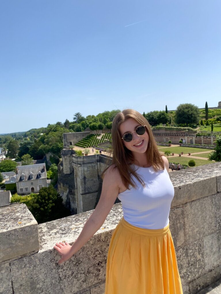
Last week, we traveled as a class to the Loire Valley. After nearly 3 hours on the bus, we finally arrived in the beautiful region of central France. Despite the heat that day, we all had a lovely time exploring the town. The Loire is the longest river in Europe, and it flows past over 20 castles. I was surprised to learn how large of a role Amboise played in the late life of Leonardo da Vinci. He spent the last three years of his life at the Château du Clos Lucé. At the château, models of his many invention ideas were on display. We have much to thank da Vinci for his contributions to our understanding of cranial anatomy, and so it was impressive to see the space in which he developed such ideas and discoveries. The gardens of the Château d’Amboise, pictured behind me are a worthy burial site for the scientific pioneer.
Last Saturday, Lauren, Sam, Rachel, another friend, and I woke up early to venture out into the countryside of France. We took a 45 minute high speed train to Reims, the city known as the unofficial capital of the Champagne wine-growing region. Many champagne houses are headquartered here, and offer tastings and cellar tours. Reims is also home to the Cathédrale Notre-Dame de Reims, a beautifully grand cathedral filled with stained-glass windows and Gothic carved portals, and has been where French kings were crowned for over 1000 years. Throughout our day of (many) champagne tastings, we tried to hone our skills and refine our palates in order to appreciate all the layers of the wines we were tasting.
Our servers commonly related the flavors of each wine with a different food. “The chardonnay offers a freshness of green apple at the beginning, and the ending flavor from the pinot noir is fruity like strawberry”. It made me wonder how in fact the intricacies of the combined biomechanics and brain mechanisms responsible for the taste come together to help us achieve the tasting of various flavors in wine. Believe it or not, there is an interdisciplinary field known as neuroenology that looks into just that.
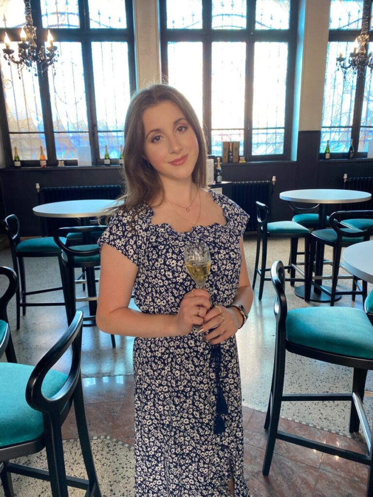
Figure 1: Me at our first wine tasting of the day. At this point we were tasting a vintage champagne from 2007.
A 2015 article by Shepherd details how this discipline investigates the contributions that science make to the enhanced quality and appreciation of wine. Many are quick to cite taste buds as the sole proprietors of flavor, however the olfactory system also plays an essential role. Olfaction begins when odorant molecules enter the nasal cavity via inhalation through the nose, or rising through the mouth from food or drink. These molecules then are able to bind to various receptors that send signals to the olfactory bulb, which activates other cascades of neural signals responsible for smell recognition, memory, and emotion (Shepard, 2015). When sniffing a glass of wine, the aromatic compounds released from the wine activate the olfactory system in a similar manner to flavor and scents previously encountered in life. In our wine tasting, we were instructed to pull a bit of air through our lips while the wine is in our mouth to aerate the wine. This further releases aromatic molecules from the wine.
Unfortunately, I cannot say we mastered the art of wine tasting by the end of the day, but the experience was truly enjoyed, nonetheless. Perhaps it is time to take up some studies on neuroenology.
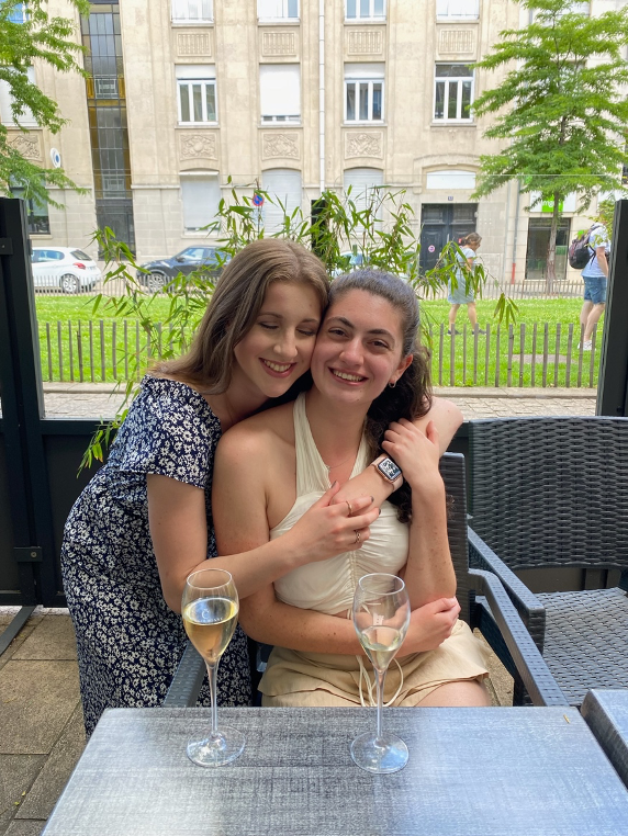
Figure 2: Clearly experiencing a boost of serotonin from the wonderful day we were having.
Reference:
Shepherd, G. M. (2016). Neuroenology: how the brain creates the taste of wine. Columbia University Press.
No trip to Paris would be complete without a visit to Museé de Louvre. The world’s most visited museum is housed in the beautiful 13th century Louvre Palace. It was fascinating to appreciate art in a space that was so illustrious, with the ceilings vying for equal attention to the paintings on the walls. Despite visiting on the day that the Mona Lisa was smeared with cake, my friend and I unfortunately did not bear witness to the protest (nor were we involved, no worries). We remained in the museum until closing, and still felt that we only scratched the surface of all there was to explore.
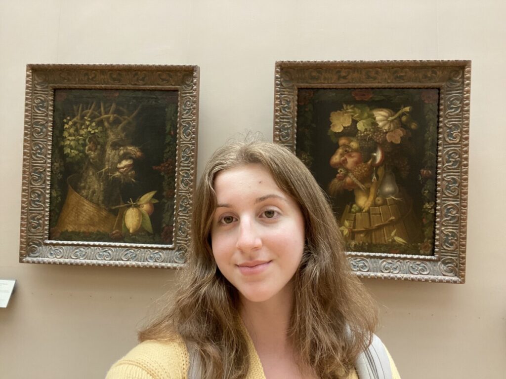
On our way out, four particular paintings caught my attention. Known collectively as The Four Seasons, these works were produced in the 14th century by Giuseppe Arcimboldo. Each one depicts a portrait composed of fruit, vegetables, and plants that relate to the respective season. Autumn represents a man but his neck is made up of pears, his chin is a pomegranate, and his ears are mushrooms. I stood there wondering why it was so familiar, and then I remembered we learned about object recognition in NBB302. Propagnosia, also known as facial blindness, is an impairment in the visual recognition of faces. Normally, faces are processed holistically but lesions in the occipital region in the ventral pathway (known as the fusiform face area) causes face blindness (Haeger et al, 2021).
Figure 1: Arcimboldo’s Autumn on display at the Louvre.
Arcimboldo’s paintings have actually been used in previous studies to observe this phenomenon. In a 2011 study, these paintings were shown to individuals with propagnosia (Rossion et al, 2011). To an unimpaired human, the elements of the painting can collectively be viewed as a face due to the object configuration. However, those with deficits in the FFA are able to only recognize the discrete components of the painting, in this case the individual fruits and vegetables. It was interesting to make this connection and it helped me more deeply understand the studies we were talking about by being able to view the stimuli in person myself.
Figure 2: Me in front of the paintings.
References:
Haeger, A., Pouzat, C., Luecken, V., N’diaye, K., Elger, C., Kennerknecht, I., … & Dinkelacker, V. (2021). Face Processing in Developmental Prosopagnosia: Altered Neural Representations in the Fusiform Face Area. Frontiers in behavioral neuroscience, 15.
Rossion, B., Dricot, L., Goebel, R., & Busigny, T. (2011). Holistic face categorization in higher order visual areas of the normal and prosopagnosic brain: toward a non-hierarchical view of face perception. Frontiers in human neuroscience, 4, 225.
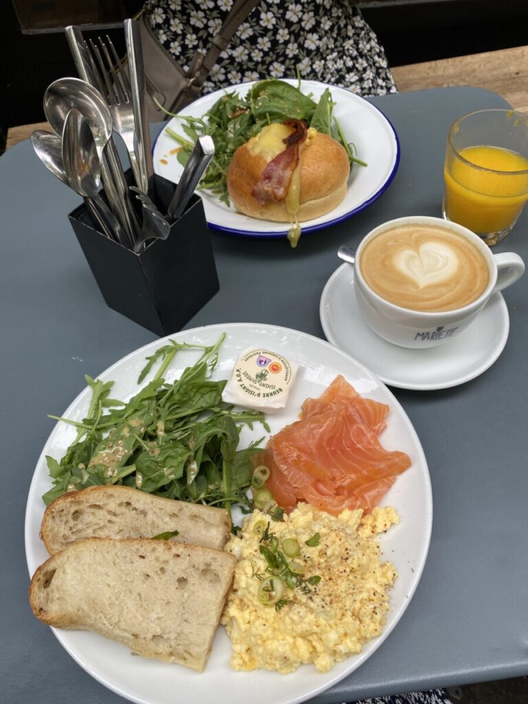
One of the best parts about coming to Paris has to be all of the delicious food I have been able to try. From a soft croissant or pain au chocolat from the local boulangerie in the morning, to fresh produce from stands and small markets dotting the streets, or a warm quiche from one of our lunch spots near Accent, I have been enjoying every tasty experience while here. The French also highly value time around meals, and so going to a cafe or restaurant is always a relaxing, serotonin-boosting time. One morning for brunch, I had a yummy plate with smoked salmon, eggs, and fresh bread. The contrasting crunch of the baguette’s crust to the warm, chewy interior was delectable. Thankfully the carbohydrates in the bread (in moderation) are as rewarding for our brain to provide it with energy as for our mouths when we taste it. 🙂
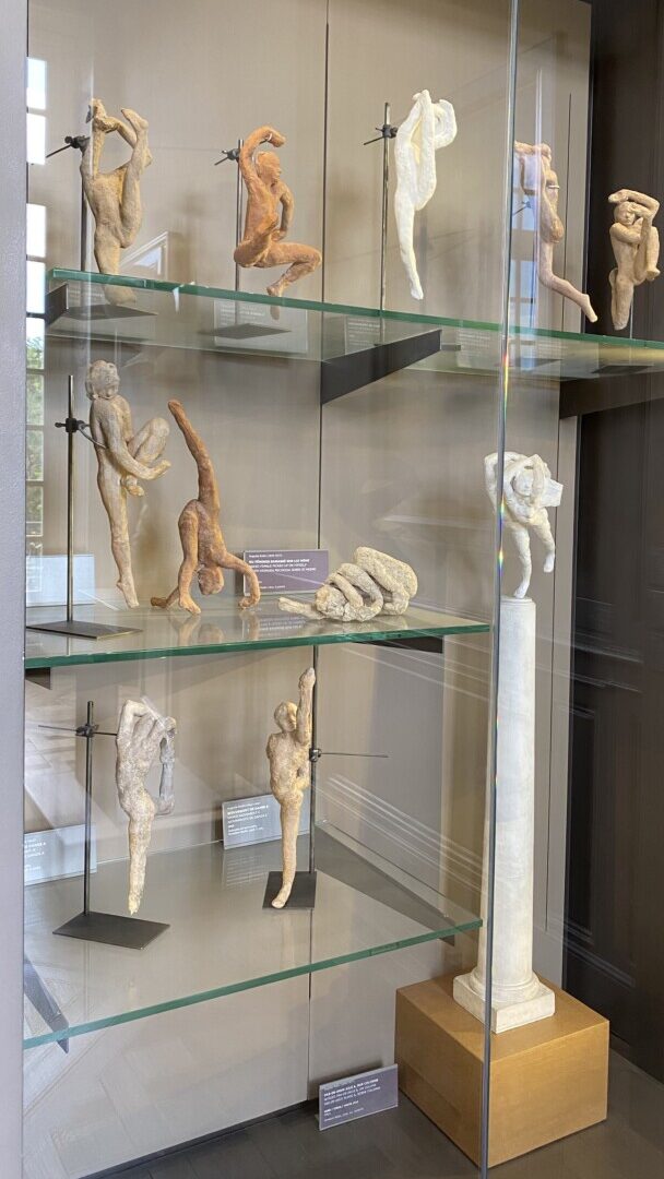
A few weeks ago when one of my friends was in town, we decided to check out the Musée Rodin. Auguste Rodin is one of my favorite sculptors, and so I was excited to visit a museum that was solely dedicated to his work. It was a wonderful experience, as it was an intimate way to explore a single artist. It is set in the Hôtel Biron, where Rodin lived toward the end of his life. Some of his sculptures are also displayed throughout the extensive garden. One of the rooms included his late sculptural experiments on dance movement studies. As a dance minor, I have always been fascinated by the link between dance and the brain. Dance demands both attention and memory skills, and significant neuronal growth seen in structures such as the insula and cingulate gyrus demonstrate the beneficial role dance can play in cognitive improvement.
One of my favorite places I have visited so far in Paris has to be The Sainte-Chapelle. It is a royal chapel in the Gothic style set within the Palais de Justice de Paris and was consecrated on April 26, 1248. It contains some of the most beautiful stained glass I have seen. The way the sun shines through the 15 large windows floods the chapel with a rainbow of color. This reminded me of learning about how the brain processes information from our eyes to perceive color. After light hits the rods and cones in the retina, the optic nerve connects to the thalamus to process those signals. The visual cortex helps us recognize what we see. I would recommend anyone visit the chapel for a highly stimulating experience!
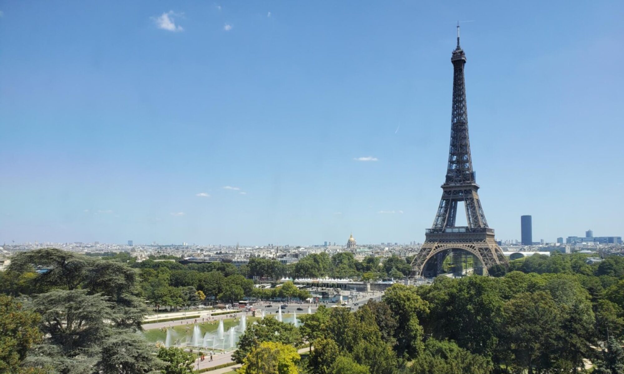
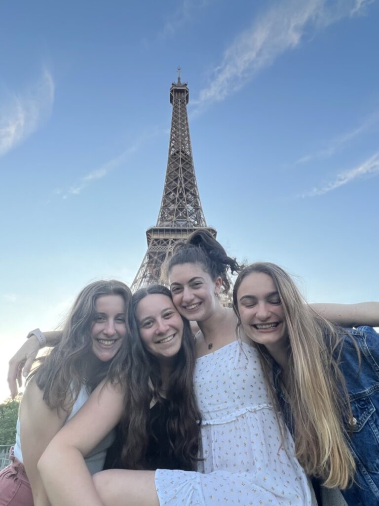
This past Wednesday, a few of us visited the Eiffel Tower for a picnic on the grass. It was a lovely evening filled with friends, food (i.e. cheese, wine, and baguettes in typical French fashion), and laughs, set underneath the iconic setting of the Eiffel Tower. We definitely were not the only ones enjoying the scenery, as the park was packed with couples, family, and friends soaking up the sun on this fine summer night.
Figure 1: Rachel, Sam, Lauren, and I in front of the Eiffel Tower during our picnic.
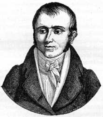
Constructed for the 1889 Worlds Fair, the wrought-iron lattice structure has become an emblem of the city and is recognized worldwide. The tower is actually the most visited monument with an entrance fee in the world! When Gustave Eiffel designed the tower, he decided to engrave the names of 72 French scientists, engineers, and mathematicians. I had never realized this scientific connection on my visits to Paris before, and began to wonder whether there were any links to neuroscience. It requires a closer look at the arch, but they are there! One such name I found is Marie François Xavier Bichat. His name appears on the first floor of the tower, 14th on the west facing side.
Figure 2: Portrait of Marie-François-Xavier Bichat. (source: https://www.wonders-of-the-world.net/Eiffel-Tower/Pantheon/Marie-Francois-Xavier-Bichat.php)
Xavier Bichat lived from 1771 to 1802, and trained in medicine in Lyon before moving to Paris. He was interested in investigating the pathology of diseases, and in 1799 he left his career as a surgeon to devote his time to experimental physiology, dissection, and autopsies to understand pathological anatomy. He is quoted to have slept in the morgue some nights in order to continue with as many dissections as possible. One of his greatest contributions was identifying and introducing 21 types of tissues as the basic elements of organs. Of even greater interest to us as neuroscience students is this pioneer’s understanding of the brain and nervous system. In his doctrine of life, he claims that the center of the animal life was the brain, and that of the organic life was the heart. For him, “life rests upon a tripod made of respiration, circulation, and nervation” (Haller 1981). He did, though, acknowledge that there is a dependent relationship between the heart and brain, in that the heart provides blood flow to stimulate cerebral tissue. In addition, he identified the importance of the ganglionic nervous system, and described it as a network of tiny, independent brains within the chest cavity. Overall, it is clear that Xavier Bichat deserves to have his name permanently inscribed on the Eiffel Tower and visible to the millions of visitors every year.
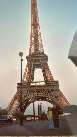
Figure 3: A fun throwback from a previous trip I had taken to Paris.
Reference:
Clarac, F., Barbara, J. G., Broussolle, E., & Poirier, J. (2012). Figures and institutions of the neurological sciences in Paris from 1800 to 1950. Introduction and Part I: Neuroanatomy. Revue neurologique, 168(1), 2-14.
Haller, J.S., 1981. American Medicine in Transition, 1840–1910. University of Illinois Press, Chicago, p. 13.
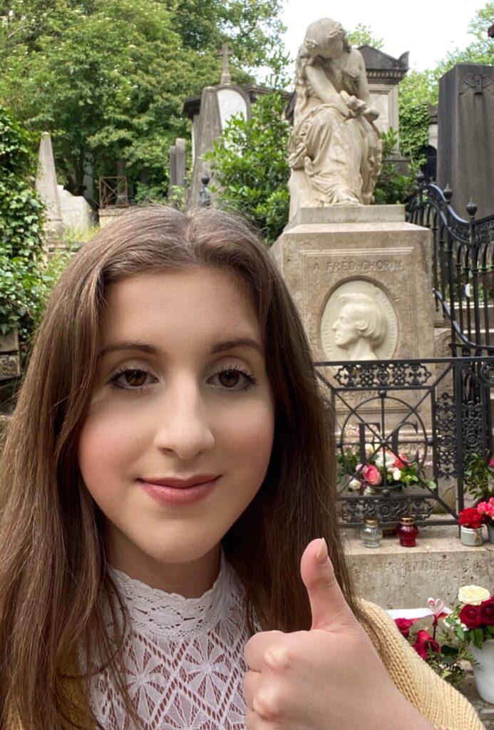
On Thursday, our class paid a visit to the Cimetiere du Pere Lachaise, a famous cemetery in Paris where many prominent figures are buried. What initially struck me was the intricacies of the burial structures, some complete with doors and stained glass. I listened to Rick Steves’ audio guide of the cemetery as I wandered through the cobblestone streets. On one track, I noticed a familiar song- The Minute Waltz, by Frédéric Chopin. It was a piece I had learned to play when I was a kid. Next thing I knew, I was standing in front of Chopin’s flower-adorned grave. Chopin had often been coined as a child prodigy. This led me to wonder whether his brain had structural differences from other non-musicians buried nearby, and if so, whether these differences were a product of nature or of nurture.
Figure 1: Me standing in front of Chopin’s grave.
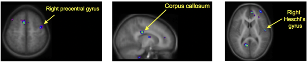
A study by Hyde et al. from 2009, titled “Musical Training Shapes Structural Brain Development”, investigates training-induced brain plasticity. It has been previously reported that adult musician brains have both structural and functional differences in musically relevant brain regions, such as auditory, sensorimotor, and multimodal integration areas, when compared to nonmusican adults. However, this study uniquely tries to answer whether there is a relationship between structural and behavioral changes in the developing brain, elucidating if structural differences in adults is a biological predisposition or a product of training at an early age. They conducted a longitudinal investigation of instrumental music training in children around age 6. Through behavioral tests and MRI scanning, the researchers found that regional structural brain plasticity only occurred in the developing brain of the instrument-training children. Before training, there were no significant differences in brain or behavior between the instrumental and control groups. By the end of the 15 months, the instrument group demonstrated significant gain in relative voxel size of the primary motor and auditory areas, and corpus callous. These were all correlated positively with behavioral improvements on motor and auditory-musical tests. This provides strong evidence that such development is induced by instrumental practice rather than pre-existing biological precursors of music ability.
Figure 2: Regions of the brain that showed plasticity following instrument training. (right precentral gyrus= primary motor area, right Heschl’s gyrus= primary auditory area). https://www.jneurosci.org/content/jneuro/29/10/3019.full.pdf
I have become interested in exploring intervention methods, such as music training, that could facilitate neuroplasticity in children with developmental disorders. Neuroplasticity is something I have not been able to explore much in other NBB courses, and thus was excited to read more about it. Having grown up practicing piano, I was curious if and how this helped shape my brain. Maybe my and Chopin’s brains are not that structurally different after all 😉
Reference:
Hyde, K. L., Lerch, J., Norton, A., Forgeard, M., Winner, E., Evans, A. C., & Schlaug, G. (2009). Musical training shapes structural brain development. Journal of Neuroscience, 29(10), 3019-3025.