A Visit With Unique Wax Models

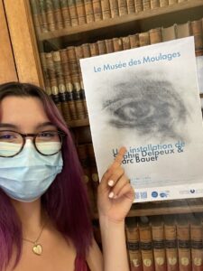
Today we ventured to the Musée de Moulages for the second time. Although it was an optional excursion, I was excited to go and see the different skin disease and conditions. Traveling to the museum allowed us to walk through a new arrondissement of Paris and see a beautiful canal. Held within hospital grounds, we saw patients enjoying the outdoors and doctors taking a break or walking to a neighboring building in the hospital.

One of the photographs displayed in the entryway of the museum to illustrate a dermatological condition.

Upon entering the museum, we saw drawings and paintings of patients with different skin conditions. In the entryway, there was a sculpture of Louis IX, the ancestor of Henry IV. Upstairs housed the real exhibit: over 4,800 castings of all types of dermatological problems, from syphilitic chancres to eczema to elephantiasis. Jules Baretta is the one who first started making the wax dermatological models after his great success with his realistic models of fruit. Despite these fascinating displays, we were strictly prohibited from taking photos, as they came from moldings of real patients, so the museum is protecting their privacy. I think it is important that the museum values the privacy of the patients so strongly, even though the patients are most likely dead.

A selfie with the different photographs and drawings of dermatological patients.

I was most fascinated by the syphilis exhibition, one of their largest displays with 442 wax models. In class, we learned about neurosyphilis and ocular syphilis and their corresponding symptoms. As I was a part of the group who presented on this target article, I had the pleasure of finding Google Images of these symptoms, such as the chancres. I realized that Google Images only displayed the mild versions of the symptoms. The extent to which syphilis can affect the body is truly horrifying, most of which I don’t think I’ll be able to unsee. I think modern medicine has come a long way since the creation of some of these castings. Doctors can detect syphilis sooner and treat it easily with penicillin, so the symptoms are not as severe.

It is important that these skin diseases are still understood and studied, even though we use photographs instead of wax models, because there has been a rise of syphilis in the past two decades, specifically in women and congenital syphilis. The museum displayed the presence of congenital syphilis in babies, and it was very unfortunate. Testing for neurosyphilis can still be difficult, but doctors normally rely on the CSF Venereal Disease Research Laboratory with a higher titer cutoff to diagnose neurosyphilis (Chow, 2021). Without modern medicine and antibiotics, a lot more people would die from syphilis and neurosyphilis.

Chow F. (2021). Neurosyphilis. Continuum (Minneapolis, Minn.)27(4), 1018–1039. https://doi.org/10.1212/CON.0000000000000982

La Trephine

The rapidity at which the field of neurosurgery has continued to evolve became evident to me during this week’s visit to Musee d’Histoire de la Medicine (Museum of the History of Medicine). We began the tour by observing what, at first, seemed to be ancient hunting tools from the San people of Southern Africa. They were mismatched in size, had asymmetrical designs, and had a mix of smooth and jagged edges. I soon found out that I was staring at the first neurosurgical toolkit used by an early French neurosurgeon. It amazed me at how these rudimentary tools were used to penetrate and tinker with an organ as complex and delicate as the human brain. Even more incredible was how these tools served as the foundation for numerous generations worth of advances in neurosurgical equipment. As we progressed down the glass boxes encasing the toolkits, I noticed them to start to garner a more sophisticated form. They became increasingly symmetrical and gradually adopted a more streamlined appearance. Interestingly, I noticed how aesthetics became more of a priority. Near the beginning of the tour, prosthetic limbs had the appearance of a robotic arm. However, closer to the 19th and 20th century, the prosthetic limb, once decked out in rusted metal, was replaced with a pale tone and easily distinguishable fingers.

Image 1: Me at soaking in the rich medical history at Musee d’Histoire de la Medicine. 

Later in the tour, I stumbled across a tool called “La Trephine” meaning “The Trepan”. In medical practice, a trepan is used to create a hole in the skull in order to expose different parts of the brain for operation. Nowadays, with more advanced technology being accessible, a cranial drill is used and is often battery-powered. The trepan in the museum, however, did not look like modern day cranial drills. It was shaped like a “Y” and had a cylindrical bottom where it attached to the brain. In order to use this tool, the surgeon would grab the top parts and twist repeatedly to make circular incisions into the skull until the bone could be removed. I discovered that this tool was commonly used to treat individuals with epilepsy. Past neuroscientists thought that mental disorders could be treated by creating an opening in the skull to allow for the demons to escape. Today, we know that epilepsy is treated with anti-epileptic drugs (AEDs) that decrease membrane excitability by interacting with neurotransmitter receptors and ion channels (Macdonald et al., 1995). This visit was very exciting and allowed me to develop a newfound understanding and appreciation for how far medicine has truly come.

Reference:

Macdonald, R. L., & Kelly, K. M. (1995). Antiepileptic drug mechanisms of action. Epilepsia, 36(s2). https://doi.org/10.1111/j.1528-1157.1995.tb05996.x

Faces of the Louvre


No trip to Paris would be complete without a visit to Museé de Louvre. The world’s most visited museum is housed in the beautiful 13th century Louvre Palace. It was fascinating to appreciate art in a space that was so illustrious, with the ceilings vying for equal attention to the paintings on the walls. Despite visiting on the day that the Mona Lisa was smeared with cake, my friend and I unfortunately did not bear witness to the protest (nor were we involved, no worries). We remained in the museum until closing, and still felt that we only scratched the surface of all there was to explore.

On our way out, four particular paintings caught my attention. Known collectively as The Four Seasons, these works were produced in the 14th century by Giuseppe Arcimboldo. Each one depicts a portrait composed of fruit, vegetables, and plants that relate to the respective season. Autumn represents a man but his neck is made up of pears, his chin is a pomegranate, and his ears are mushrooms. I stood there wondering why it was so familiar, and then I remembered we learned about object recognition in NBB302. Propagnosia, also known as facial blindness, is an impairment in the visual recognition of faces. Normally, faces are processed holistically but lesions in the occipital region in the ventral pathway (known as the fusiform face area) causes face blindness (Haeger et al, 2021).

Figure 1: Arcimboldo’s Autumn on display at the Louvre.

Arcimboldo’s paintings have actually been used in previous studies to observe this phenomenon. In a 2011 study, these paintings were shown to individuals with propagnosia (Rossion et al, 2011). To an unimpaired human, the elements of the painting can collectively be viewed as a face due to the object configuration. However, those with deficits in the FFA are able to only recognize the discrete components of the painting, in this case the individual fruits and vegetables. It was interesting to make this connection and it helped me more deeply understand the studies we were talking about by being able to view the stimuli in person myself.

Figure 2: Me in front of the paintings.

References:

Haeger, A., Pouzat, C., Luecken, V., N’diaye, K., Elger, C., Kennerknecht, I., … & Dinkelacker, V. (2021). Face Processing in Developmental Prosopagnosia: Altered Neural Representations in the Fusiform Face Area. Frontiers in behavioral neuroscience15.

Rossion, B., Dricot, L., Goebel, R., & Busigny, T. (2011). Holistic face categorization in higher order visual areas of the normal and prosopagnosic brain: toward a non-hierarchical view of face perception. Frontiers in human neuroscience4, 225.

Ballet: A Workout for Brain and Body

Me in my best attempt at a Versailles-esque outfit standing outside the gates to the palace after the ballet performance.

The last time I was in Paris, it was for ballet class when I was in the eighth grade. Through the language barrier and intimidation factor of taking a class in a new country, I loved my ballet studio here and the opportunity to connect with girls I couldn’t directly communicate with through an art form we all loved. So, this time when I touched down in Paris, I knew that I wanted to find a way to appreciate the thing that brought me here in the first place. After researching with my roommates, we discovered that we would be in Paris over the 250th anniversary of Marie Antoinette’s wedding and that it was being celebrated with a ballet at the Versailles Royal Opera House. So, on a rainy weekend day, we put on our most Versailles-appropriate dresses and made the hour and a half-long journey to the Opera House. 


The inside of the Opera House before the show; this is the same Opera House that Marie Antoinette watched performances in while she lived at Versailles during her husband’s rule.

The ballet itself was incredible, with historical context provided on a screen above the stage and the dancers carefully telling the story of Marie Antoinette’s life, death, and antics on stage. Amidst the colorful costumes, contemporary take on classical ballet, and endless violin swells, I felt reconnected to my love for dance; the opportunity to experience that same passion in such a unique way was beyond incredible, and I know that the memory of that night is one I’ll cherish forever. 

Connecting this experience back to neuroscience, though, was equally as interesting. Having grown up dancing my whole life, I knew there were lessons I’d learned and habits I’d built that stick with me even today (I still find myself measuring eight-counts in music and counting steps as I walk across a room). But, in light of the courses we’re taking here, I thought it would be interesting to explore the connection between dance and the brain. Through my research, I found an interesting study which explored the relationship between dance and neural plasticity. Specifically, researchers observed structural changes in the hippocampal and grey-matter volume of dancers, as well as heightened levels of white-matter function. These physical differences then led to functional improvements in memory, body balance, and several other realms. The researchers thus concluded that dance can, in fact, improve neuroplasticity by integrating brain regions (Teixera-Machado et al, 2019). 

The dancers and orchestra taking their final bows after the performance.

Overall, I think that the opportunity to connect the purposes behind both my visits to Paris was so special, and I’m glad that I had the opportunity to find a way to bring my time here full circle. I know that being here has confirmed my love for both neuroscience and dance, and I am already researching dance studios at home to continue exploring my passions from an ever-increasingly interdisciplinary perspective. 


References:

​​Teixeira-Machado, L., Arida, R. M., & de Jesus Mari, J. (2019). Dance for neuroplasticity: A descriptive systematic review. Neuroscience and biobehavioral reviews, 96, 232–240. https://doi.org/10.1016/j.neubiorev.2018.12.010


Compasses, calculators, and robots, oh my!

Compasses, calculators, and robots, oh my! Today, we attended the Musée des Arts et Métiers to learn all about scientific instruments throughout the ages.  One of my favorite things in the museum was a device from the late 18th century used to ferment wine. Wine fermentation is truly an art form and has taken centuries to perfect into modern wine we drink from a bottle today and getting to see an original fermentation device was such an intriguing experience.

Me standing in front of a grandfather clock after viewing the medical device portion of the museum


We also got to view a magnetic drum device, a precursor to the modern calculator we use today. It strangely resembled a typewriter. We also viewed different communication devices from years (and even centuries) past, including the first press-cylinder printer and an original Nokia from the early 2000s. Additionally, we saw a Russian extra-terrestrial rover that could communicate from outer space! Lastly, my favorite thing we got to see was a thermoscope from 1592 that once belonged to Galileo! In fact, with some further research after leaving the museum, I discovered that Galileo actually introduced the first thermoscope thermometry which would eventually evolve to the modern thermal imagery we see today!

Galileo’s original thermoscope from 1592


Evidently, this invention sprung medicinal science forward towards modern Western biomedicine. Fever has always been one of the most common medical indicators of ailment, and it made me ponder how temperature correlates with neuroscience and potential brain injuries. I found a recent study that discussed the relationship between body and brain temperature in rodents with traumatic brain injuries (TBIs). Previous research had previously indicated that extreme temperature deficits or increases may affect the implications of brain injuries. Researchers decided to compared rats that had acquired traumatic brain injuries to uninjured rats both under general anesthesia and not under general anesthesia. When under anesthesia, there was no significant temperature difference between rats with TBIs and rats without TBIs. However, rats who were under anesthesia had significantly lower temperatures than rats not under anesthesia, suggesting that anesthesia alone caused a decrease in temperature. When rats had not been put under anesthesia temporalis muscle temperature correlated well with brain temperature, but rats with TBIs and rats without TBIs did not differ in temperature. This allowed the researchers to conclude that temporalis muscle temperature is a good indicator of brain temperature, however, brain temperature itself is not necessarily indicative of a TBI. After reading this article, it was very intriguing to consider how far the science of temperature in medicine has come since the days of Galileo.

References: 

Jiang, J. Y., Lyeth, B. G., Clifton, G. L., Jenkins, L. W., Hamm, R. J., & Hayes, R. L. (1991). Relationship between body and brain temperature in traumatically brain-injured rodents. Journal of Neurosurgery, 74(3), 492–496. https://doi.org/10.3171/jns.1991.74.3.0492

Ring, E. F. J. (2007). The historical development of temperature measurement in medicine. Infrared Physics & Technology, 49(3), 297–301. https://doi.org/10.1016/j.infrared.2006.06.029


A Tough Loss (on all accounts)

By Ally Grubman

Last Friday the class took a trip to a French Rugby match, or so we thought. About 2 minutes into the game, there were comments flying around our NBB section like “Are we sure this is rugby?”, “When are they going to pick up the ball??”, and my personal favorite, “Guys, they’re just warming up with soccer, don’t worry.” Yep, you guessed it, we made our way to the France vs. Denmark fútbol (soccer) game. So awkward for us.

At the beginning of the soccer game, before the big revelation.

But even though we went thinking it was a rugby game, there was still some relevance to our class that gave me further insight into what we’ve been learning. In our NBB402W class last week, we read a paper on rugby players and the correlation between concussions and later cognition and neurodegenerative illnesses. Although we didn’t get to actually see any rugby players get head injuries, we did however see lots of soccer players use their heads to hit the ball. While this might not be as aggressive or harmful as rugby, there’s no way it’s good for you. For instance, Broglio et al. (2003) found that over a 300-game career, a soccer player experiences roughly 2000 headers, which can add up to cause a lot of damage. This only includes games, not even considering those sustained during practice. This repeated injury can lead to real problems in the future, specifically neurodegeneration. When I say neurodegeneration and neurodegenerative diseases, think: Alzheimer’s, ALS, and Parkinson’s (and so many more). This is something we also found in our rugby paper. Rugby players are rarely required to wear protective or effective helmets. Additionally, since it is very much a contact sport, there has been an increased need for research on the implications of playing such a dangerous game. Tomasin et al. (1989) outline the importance of more awareness on the topic, specifically concerning rugby players and the risk they are putting themselves in. Coaches, physicians, and players need to be more aware of the dangers and the serious possibility of brain damage and degeneration in the player’s future.

A group selfie at the beginning of the game to commemorate the fun time we all had!

However, while we counted the headers and enjoyed the game, we had a great time hanging out in a non-school setting, eating some very overpriced stadium food, and laughing about the fact that we were, in fact, not at a rugby game. It’s too bad that France lost, but all in all, it was a great game and a ton of fun for all of us!!

Citations:

Broglio, S. P., Ju, Y. Y., Broglio, M. D., & Sell, T. C. (2003). The Efficacy of Soccer Headgear. Journal of athletic training, 38(3), 220–224. 

Tomasin, J. D., Martin, D. F., & Curl, W. W. (1989). Recognition and Prevention of Rugby Injuries. The Physician and sportsmedicine, 17(6), 114–126. https://doi.org/10.1080/00913847.1989.11709809

Wonderful carbs at Cafe Marlette (Photo post)

One of the best parts about coming to Paris has to be all of the delicious food I have been able to try. From a soft croissant or pain au chocolat from the local boulangerie in the morning, to fresh produce from stands and small markets dotting the streets, or a warm quiche from one of our lunch spots near Accent, I have been enjoying every tasty experience while here. The French also highly value time around meals, and so going to a cafe or restaurant is always a relaxing, serotonin-boosting time. One morning for brunch, I had a yummy plate with smoked salmon, eggs, and fresh bread. The contrasting crunch of the baguette’s crust to the warm, chewy interior was delectable. Thankfully the carbohydrates in the bread (in moderation) are as rewarding for our brain to provide it with energy as for our mouths when we taste it. 🙂

Artist in Motion– and stillness (Photo post)

A few weeks ago when one of my friends was in town, we decided to check out the Musée Rodin. Auguste Rodin is one of my favorite sculptors, and so I was excited to visit a museum that was solely dedicated to his work. It was a wonderful experience, as it was an intimate way to explore a single artist. It is set in the Hôtel Biron, where Rodin lived toward the end of his life. Some of his sculptures are also displayed throughout the extensive garden. One of the rooms included his late sculptural experiments on dance movement studies. As a dance minor, I have always been fascinated by the link between dance and the brain. Dance demands both attention and memory skills, and significant neuronal growth seen in structures such as the insula and cingulate gyrus demonstrate the beneficial role dance can play in cognitive improvement.

Musee de Moulages: the skin-brain axis

This Tuesday, June 7th, we had the opportunity to visit the Musee de Moulages at the Hospital Saint-Louis. The museum houses “a unique collection of wax dermatological models”, as coined by the subtitle on the front cover of the museum pamphlet.

Pictured above is the museum pamphlet containing interesting historical context about their collection and dermatology as a practice!

Reflecting upon my visit, I can concur that this museum has a unique collection indeed. With collections of wax models displaying pathologies such as syphilis, tuberculosis, eczema, acne, elephantiasis, tumors, and much more, the museum leaves a lasting impression. 

Me in front of the museum flyer 🙂

The first week of the program, we learned about the “gut-brain axis” with the first target article. Upon visiting the dermatology museum, I began to wonder if the same kind of connection exists between the brain and the skin, a “skin-brain axis”. Considering that the skin is the largest organ in the body, I postulated that this connection must exist in some form or another. Before diving into the current scientific literature, I speculated on different relationships that might exist between the brain and the skin: How do neurological variables such as stress manifest in the skin? How does skin pathology affect the brain? What kinds of information does the skin communicate to the brain and through what mechanisms?

I found that the “brain-skin” connection does exist, and lends itself to a great intersection of research involving different fields of study such as psychology, neurobiology, dermatology, immunology, etc. Stress and skin conditions have been known to be associated for centuries, and modern research has established the existence of a causal relationship as psychological stress can lead to the onset and/or aggravation of multiple skin diseases. Many studies also reveal that stress accelerates skin aging (Lee et al., 2020; Rinnerthaler et al., 2015) and exacerbates skin inflammation (Chen et al., 2014). Not surprisingly, anxiety has been proven to increase susceptibility to skin diseases which one study claims is due to the disruption of cutaneous homeostasis via prolonged sympathetic nervous system activation (Öksüz et al., 2020). In addition, the relationship between depression and chronic wounds has been investigated as these conditions often have a high comorbidity (Hadian et al., 2020). 

However, in my opinion, the most interesting finding of my literature review of the skin-brain axis concerned the affective function of the skin. The discriminative properties of the skin have been well identified, however, the discovery of a new class of nerve fiber, unmyelinated C-fiber afferents, has led to a new understanding of skin function as a coder of emotional touch. C-tactile mediated touch such as massage therapy has exciting implications for clinical treatments of both physical and psychological symptoms of chronic skin conditions like eczema. 

All in all, neuroscience is a wonderfully interdisciplinary subject that has interesting implications on nearly all areas of biology and different clinical practices – including dermatology!

Chen, Y., & Lyga, J. (2014). Brain-skin connection: stress, inflammation and skin aging. Inflammation & allergy drug targets, 13(3), 177–190. https://doi.org/10.2174/1871528113666140522104422

Hadian, Y., Fregoso, D., Nguyen, C., Bagood, M. D., Dahle, S. E., Gareau, M. G., & Isseroff, R. R. (2020). Microbiome-skin-brain axis: A novel paradigm for cutaneous wounds. Wound repair and regeneration : official publication of the Wound Healing Society [and] the European Tissue Repair Society, 28(3), 282–292. https://doi.org/10.1111/wrr.12800

Lee, C. M., Watson, R., & Kleyn, C. E. (2020). The impact of perceived stress on skin ageing. Journal of the European Academy of Dermatology and Venereology : JEADV, 34(1), 54–58. https://doi.org/10.1111/jdv.15865

Lloyd, D. M., McGlone, F. P., & Yosipovitch, G. (2015). Somatosensory pleasure circuit: from skin to brain and back. Experimental dermatology, 24(5), 321–324. https://doi.org/10.1111/exd.12639 

Öksüz, Ö., Günver, G., Oba, M. Ç., & Arıkan, K. (2020). Psychiatry to dermatology; panic disorder. Journal of clinical neuroscience : official journal of the Neurosurgical Society of Australasia, 81, 316–320. https://doi.org/10.1016/j.jocn.2020.09.071 

Rinnerthaler, M., Bischof, J., Streubel, M. K., Trost, A., & Richter, K. (2015). Oxidative stress in aging human skin. Biomolecules, 5(2), 545–589. https://doi.org/10.3390/biom5020545


Seeing the Light (Photo Post)

One of my favorite places I have visited so far in Paris has to be The Sainte-Chapelle. It is a royal chapel in the Gothic style set within the Palais de Justice de Paris and was consecrated on April 26, 1248. It contains some of the most beautiful stained glass I have seen. The way the sun shines through the 15 large windows floods the chapel with a rainbow of color. This reminded me of learning about how the brain processes information from our eyes to perceive color. After light hits the rods and cones in the retina, the optic nerve connects to the thalamus to process those signals. The visual cortex helps us recognize what we see. I would recommend anyone visit the chapel for a highly stimulating experience!