The Fruits of Jules Baretta’s Labor: The Story Behind the Museé des Moulages

by Samantha Feingold

During our class visit to the Museé des Moulages, I learned about both the science and the history behind dermatology. The Museé des Moulages translates to the museum of castings and is a collection of wax models that depict the dermatological presentations of neurological diseases, including those that we have discussed in class such as syphilis (Lynn et al., 2004; Rasoldier et al., 2020). The museum had an in-depth collection demonstrating the range of skin presentations and the changing appearances throughout the four progressing stages of syphilis.
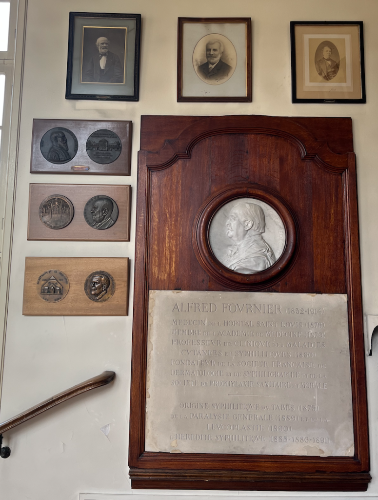
 

Figure 1. A plaque in the Museé des Moulages regarding the history of syphilis and progression of medicine, photographed by Samantha Feingold.

The story of Museé des Moulages begins with artist Jean Baretta creating models of apples in 1863. Discovered by Charles Lailler, it was suggested that Baretta use his talent to reproduce skin diseases. As a result, the first wax dermatological model was made by Baretta in 1867.

Prior to the creations of castings when photographs were not available, students depended on engravings and drawings to learn about these pathologies. Thus, it was very difficult to understand these diseases and be prepared to diagnose patients. At the Museé des Moulages, castings were made of patient skin conditions and affected body parts to aid the improvement of education and diagnosis accuracy. We were not permitted to take photographs of the castings out of respect for privacy and patient confidentiality. The castings are painted, appear incredibly realistic, and include facial and genital castings which contribute to the seriousness of privacy and respect for those patients. These unique three-dimensional models were very valuable and have curated a historical collection of de-identified patient cases. Many of the castings were quite graphic and demonstrate how far medicine has come. When these castings were made, the treatments for these disorders were nothing compared to today and those individuals’ contribution to the creation of these wax models was essential for the identification of the same diseases that can now be treated. While some of these castings were hard to look at knowing that people suffered tremendously, it is informative to see the severity of syphilis progression and many other dermatological diseases due to the inability to provide medical intervention at that time. I am grateful we had the experience to observe incredibly accurate models of these diseases that we previously were googling to understand. I am glad I now know about the history of advancing dermatology. Knowing wax molds of apples inspired this museum, I wonder if that is where the saying “an apple a day keeps the doctor away” originated from.
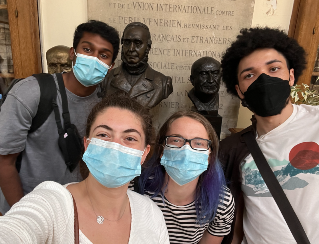
Figure 2. Adway, Alyssa, Duke, and myself at Museé des Moulages.

Works Cited

Lynn, W. A., & Lightman, S. (2004). Syphilis and HIV: a dangerous combination. The Lancet. Infectious diseases4(7), 456–466. https://doi.org/10.1016/S1473-3099(04)01061-8

Rasoldier, V., Gueudry, J., Chapuzet, C., Bodaghi, B., Muraine, M., Tubiana, R., Paris, L., Pestel-Caron, M., Caron, F., & Caumes, E. (2021). Early symptomatic neurosyphilis and ocular syphilis: A comparative study between HIV-positive and HIV-negative patients. Infectious Diseases Now, 51(4), 351–356. https://doi.org/10.1016/j.medmal.2020.10.016
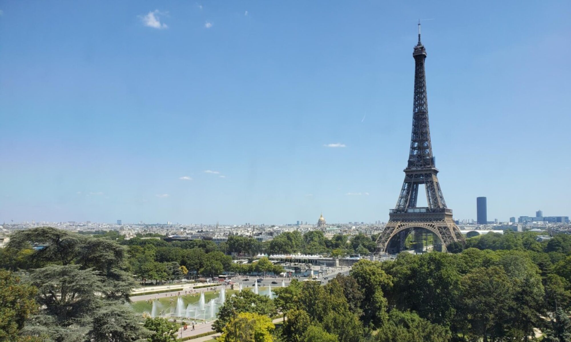
 

Baguettes, Wine, Cheese, and the Eiffel Tower (Photo Post)

When I think of Paris, the first thing that comes to mind is the Eiffel Tower. This photo shows me and Rachel on the lawn of the Eiffel Tower while we had a picnic and photoshoot with some of our classmates, sharing a baguette, cheese, and wine to celebrate our budding friendship. Although I was not sure how much I enjoyed the architecture at first, spending time in the city has allowed the tower to grow on me. The Eiffel Tower was built by Gustave Eiffel for the Paris World Fair in 1889 and features featuring the names of 72 scientists on the borders of the sides of the tower as a homage to the men of science. Despite its fame, the Parisians have not always been thrilled by its appearance, regarding the tower as an eyesore and calling for its removal until the addition of a radio tower at the top in 1909.

Stepping into the Lab of Marie Curie

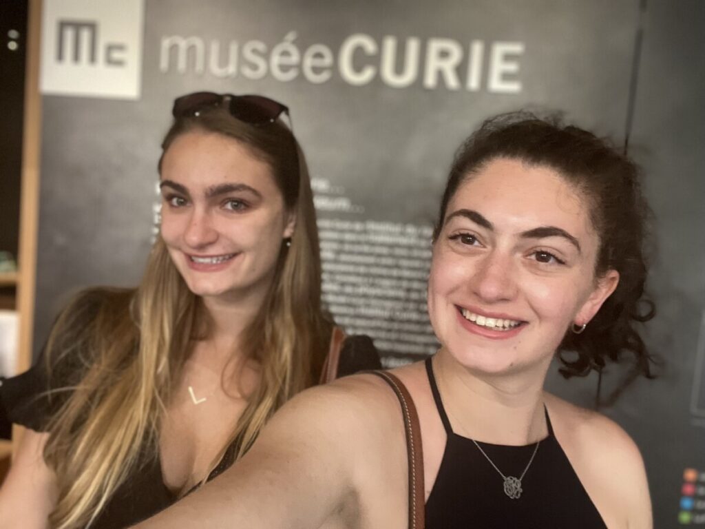
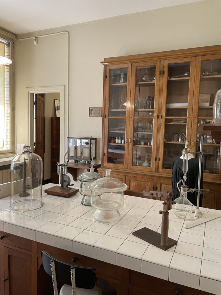
On June 15th, 2022, Cynthia, Sam, Alyssa, and I visited the Marie Curie Museum, located down the street from the Pantheon, where Marie and Pierre Curie are both buried. I was very excited to go to this museum and have been talking about it for the past few weeks because of the importance of Curie’s work in the field of oncology. The museum is located at the former Radium Institute, now called the Institut Curie, and featured Curie’s (decontaminated) lab, office, and garden. Curie spent the last 20 years of her life as the first director of Curie Laboratory at the Radium Institute, where she ensured her lab comprised of 25% of the women, before passing away from leukemia in 1934. I noticed Curie’s lab was smaller than most labs I have seen at Emory or during my research at Children’s Hospital of Philadelphia, which begs the question of whether things in the United States are just larger or if labs in the past were built smaller. The museum displayed a replication of the 1902 lab book entry by Marie and Pierre Curie estimating the atomic mass of Radium and outlining the various steps of their calculations. It’s insane to think that the original page is still dangerously radioactive over 100 years later.

Figure 1. Sam and I in the Marie Curie Museum

Marie Curie was a Polish and nationalized-French physicist and chemist who won two Nobel Prizes for her work with radioactivity. She developed contact curiethérapie, known as brachytherapy in English, a technique still used today for the treatment of cancer. Brachytherapy can be used for the treatment of a variety of cancers, including prostate, cervix, breast, vagina, endometrium, and head and neck cancers and has been shown to be an effective and safe non-pharmaceutical treatment with fewer serious complications and better outcome than other treatments for breast cancer (Deng et al., 2017). Brachytherapy has also been found to treat brain tumors with high doses of radiation while sparing the healthy surrounding tissue and can be used as a primary treatment, an adjuvant treatment, or as therapy for recurrence of some malignant gliomas, low-grade astrocytomas, meningiomas, metastases, and pediatric brain tumors (Suh et al., 1999).

Figure 2. Photograph of Marie Curie’s lab, taken at the Marie Curie Museum

I find it amazing that, even after almost a century, the science Marie Curie lived (and died) for continues to help patients around the world. She truly is an inspiration to all young girls interested in science and medicine. It was a wonderful experience to step back into the past and learn about radiation, cancer research, and the life of Marie Curie.

Citations:

Deng, X., Wu, H., Gao, F., Su, Y., Li, Q., Liu, S., & Cai, J. (2017). Brachytherapy in the treatment of breast cancer. International journal of clinical oncology22(4), 641–650. https://doi.org/10.1007/s10147-017-1155-5

Suh, J. H., & Barnett, G. H. (1999). Brachytherapy for brain tumor. Hematology/oncology clinics of North America13(3), 635–ix. https://doi.org/10.1016/s0889-8588(05)70080-0

Blogging about Braille

It is crazy to think that four weeks have already passed, only leaving us one more week in Paris. With still so many places to explore, I am not ready to say goodbye yet. However, yesterday, Wednesday, June 15th, I had the opportunity to check an item off of my bucket list when our class visited the Pantheon. As I was living very close to this monument during the first three weeks of the trip, I was very excited to finally be able to go inside. The Pantheon, originally built to be used as a church and modeled after the Roman Pantheon, features very tall ceilings with paintings that date back to the nineteenth century. In the center of the main floor is Foucault’s Pendulum, which was, unfortunately, being cleaned during our visit, so we weren’t able to see its full swing. 

Jewel, Khushi, and me inside the Pantheon crypt

After exploring the ground floor, we delved into the basement to visit the Pantheon crypt. Prior to the visit, I had a few stops that I wanted to make, such as the tomb of Marie Curie as she was the first woman to be buried in the Pantheon. However, while looking around, I was surprised to find that Louis Braille had been buried there as well. He was publicly recognized by the French government due to his creation of the Braille alphabetic writing system, which greatly contributed to the blind and visually impaired communities around the world, and his remains were taken to the Pantheon in 1952. 

The tomb of Louis Braille in the Pantheon.

There are around six million blind people around the world using Braille, stressing the importance of doing more research on it. Reich, Szwed, Cohen, & Amedi 2011 explored the areas of the brain that could potentially play the role of the visual word form area (VWFA) in people with visual impairment. They recruited eight congenitally blind individuals to partake in multiple different experimental conditions: braille words, braille nonsense words, verb generation, and verb generation control. Their participant results and associated functional magnetic resonance images (fMRI) were compared to data from Cohen et al., 2004 to highlight that visual word form area activation occurred almost identically in visually impaired individuals and sighted, suggesting a “specialization for reading regardless of visual experience”. It is fascinating to learn that the contributions of one man, Louis Braille, have allowed visually impaired individuals to read, even though reading is typically thought of as a unique visual experience. 


References:

Reich, L., Szwed, M., Cohen, L., & Amedi, A. (2011). A Ventral Visual Stream Reading Center Independent of Visual Experience. Current Biology, 21(5), 363–368. https://doi.org/10.1016/j.cub.2011.01.040

Cohen, L., Jobert, A., Le Bihan, D., & Dehaene, S. (2004). Distinct unimodal and multimodal regions for word processing in the left temporal cortex. NeuroImage, 23(4), 1256–1270. https://doi.org/10.1016/j.neuroimage.2004.07.052

12 hours at the Eiffel Tower!


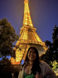
Me and the Eiffel Tower!


Following a busy week of assignments– two papers and a small presentation later– my roommate and I were ready for the weekend! Before arriving in Paris, Solanch, Jewel, and I booked a tour of the Eiffel tower and it turned out to be the perfect adventure after a long week. During the first three weeks in Paris, Solanch and I enjoyed seeing the Eiffel tower twinkle from our apartment kitchen each night before bed. We knew we wanted to enjoy the beauty of the tower as much as possible, so we decided that we would take full advantage of our visit and spend the entire day in the area. While we have gotten the chance to study at the accent center, the nearby cafes, and even the Sainte-Geneviève library, we wanted to experience studying with the tower in the background. We decided that the view would inspire us as we worked on our assignments so we brought our computers to get ahead on some readings. 

Some of the names along the Eiffel Tower.


As we walked toward the tower, we learned that Gustave Eiffel’s apartment could be found at the top of the tower. Apparently, the space was used to accommodate his guests and work on scientific experiments– how cool is this? As I heard this, I thought to myself: I wouldn’t mind the commute every morning! Before reaching the lines to start our ascension, we could already see the Eiffel tower in all of its glory. It towered 1083 feet above us and was even more breathtaking up close. As we got closer and closer, our tour guide pointed out the names that bordered the four sides of the tower. Apparently, the names of 72 scientists, including mathematicians and engineers, were inscribed along the four sides of the tower. I wondered if there were any neuroscientists that had made the cut. A quick google search told me that Marie-François Xavier Bichat, although not a neuroscientist by training, was an anatomist who made substantial contributions that were later used for the development of neuroscientific understanding. He is most recognized for establishing the Medical Society of Emulation and for his publications titled: Physiological Researches on Life and Death and General Anatomy Applied to Physiology and Medicine (Clara et al., 2012). Through many***  dissections of corpses, he was able to identify 21 different types of elementary tissues. All of this was done without the use of a microscope! Pretty remarkable! I love how history is so integrated and evident all throughout Paris. I can’t believe that we have a little over 1 week left of this incredible experience!

Me and Eiffel Tower at night!


Clarac, F., Barbara, J.-G. ., Broussolle, E., & Poirier, J. (2012). Figures and institutions of the neurological sciences in Paris from 1800 to 1950. Introduction and Part I: Neuroanatomy. Revue Neurologique, 168(1), 2–14. https://doi.org/10.1016/j.neurol.2011.08.013


NBB 201 as a Museum: Visiting Musee de l’Homme

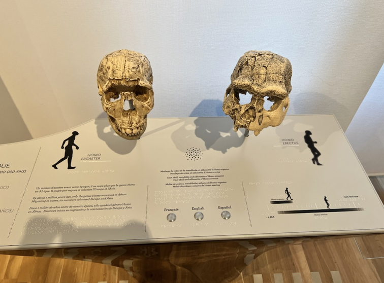
Skulls of a Homo erectus and Homo ergaster.

On Monday, June 13th, the class took a visit to Musee de l’Homme, an anthropology museum. This museum focused on what makes humans unique from other species and how we evolved. There was a display showing brains of different species emphasizing the size differences. One of the brains on display was rhesus macaques, the type of monkey I work with in my lab. Even though I have worked with the monkeys and seen how small their heads are, I didn’t realize how small their brains are because on my computer they look so big! Something else I really enjoyed about the museum is how much of hominin evolution was familiar to me from NBB 201. For example, pictured in this post are skulls of a Homo erectus and Homo ergaster, both species I learned about. It was really nice to actually see what I have learned.

What would your pen name be?

Today (Wednesday), the class visited the Pantheon in Paris. I had no idea that such a building existed, and I was very surprised to know that a select few were buried in the crypts beneath it.

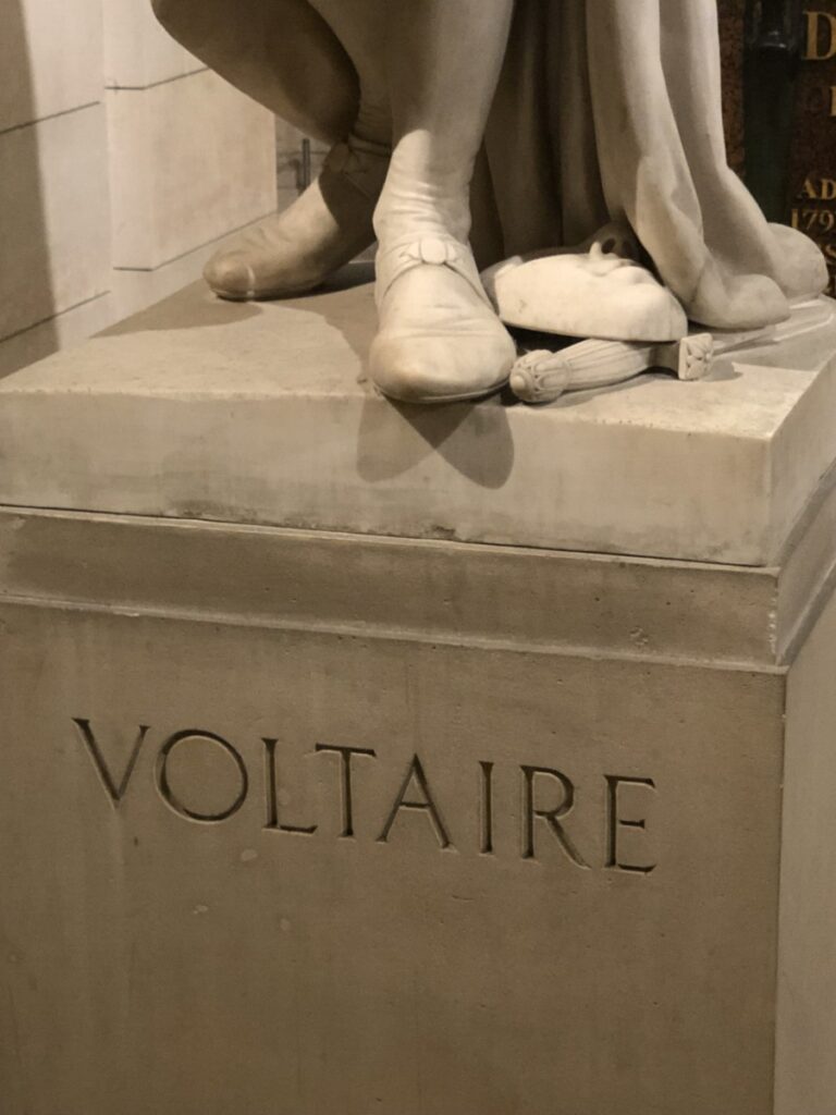
Personally, the big name for me was François-Marie Arouet, known by his nom de plume (pen name) Voltaire. In my preparation to come to Paris, I read his book, “Candide,: which turned out to be a very readible story that I might even compare to a text such as “The Alchemist” by Paulo Coelho. 

This is a statue of the famous philosopher of the pen name, Voltaire. Very cool.

I wonder, however, whether any of the folks buried in the crypt would even consent to being there–it seems that these “important” people were moved posthumously. Perhaps this was a disservice.

The list of 70 figures buried in the crypt include Rosseau, Curie, and even Braille. 

I did not know very much about Marie Curie prior to the visit, and I found that there was actually a pubmed article dedicated to her:

“Marie Curie was a remarkable woman whose discoveries broke new ground in physics and chemistry and also opened the door for advances in engineering, biology, and medicine. She broke new ground for women in science: she was, for example, the first woman to receive a doctor of science degree in France, the first woman to win Nobel Prize, the first woman to lecture at the Sorbonne, the first person to win two Nobel Prizes, and the first Nobel Laureate whose child also won a Nobel Prize. Her life offers insights into the changing role of women in science and academia over the past century. It also offers examples of many ways in which scientists can, and should, work to improve the educational programs and career opportunities available to those who follow in their footsteps.”

However, in response to the prompt offered by the NBB 402W rubric on the question:

“Marie Curie is one of the only women at the Pantheon. Why?”

I felt it would be more appropriate to talk about a female scientist who was not celebrated and honored due to the systemic imbalance of power she faced. In our NBB 471 class, our guest speaker, Dr. Herve Chneiweiss, made several references to Dr. James Watson. Even in the context of this discussion, there was no credit given to Rosalind Franklin who actually discovered the “informative X-ray diffraction patterns of DNA that provided vital clues for James Watson and Francis Crick’s double-stranded helical model.” 

Rather, she was overshadowed by the sexism in science, back when DNA was discovered, and today, in class when she was not a part of the conversation of genetic research. She continued her research and maintained correspondence with Watson and Crick through her study of diffraction patterns of Tobacco mosaic virus–details all swept under the rug by very apparent sexism everywhere we look. 

To answer the question of “why” the one-word answer is: sexism.

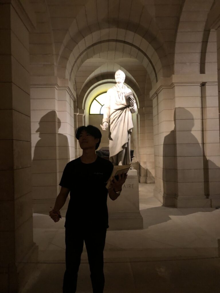
This is a picture of me being Voltaire. He actually satirizes sexism in the book that I read, Candide. I would recommend it. Go Voltaire.

p.s. my last Parker pen, a gift from a dear high school friend, was stolen on a trip to Barcelona, and so i purchased a Caran d’Ache pen from Palais du Stylo. i would highly recommend a visit here if you are interested in a new writing utensil. also, a very fun fact is that is that

“Caran d’Ache was the pen name for Emmanuel Poiré (6 November 1858 – 25 February 1909). The pseudonym comes from Russian: карандаш, romanized: karandash meaning “pencil” in Turkic languages. While his first work glorified the Napoleonic era, he went on to create “stories without words” and as a contributor to newspapers such as the Le Figaro, he is sometimes hailed as one of the precursors of comic strips. The Swiss art products company Caran d’Ache is named after him”

My precious.

this fun post-script fact brings us to a full circle ending as Voltaire was a pen name as well.

voila.

References:

https://pubmed.ncbi.nlm.nih.gov/15482656/

https://pubmed.ncbi.nlm.nih.gov/18702397/

https://en.wikipedia.org/wiki/Caran_d%27Ache

I see dead people…in crypts


I see dead people…in crypts. On Wednesday, we went to visit the Pantheon in Paris! “Pantheon” is derived from Greek, meaning ‘temple for all gods.’ However, the French Pantheon was intended not to be a religious symbol or house any religious artifacts. Instead, this Pantheon would be the resting place for those who extended the greatness of France, and assisted France in pursuing its’ national motto: liberté, égalité, fraternité (liberty, equality, and brotherhood.)

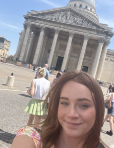
Me in front of the Pantheon when we first arrived

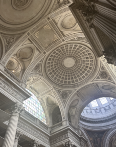
The ceiling of the Pantheon features high arches


While listening to the audio tour, I learned that only men (and, more recently, women) who made their achievements after July 14, 1789, are allowed to be buried at the Pantheon. This is because only those who brought glory to France during ‘freedom’ may be buried there, and July 14, 1789, was the Storming of the Bastille that was recognized as the official beginning of the French Revolution, and every period after that was determined to be free France. This can be seen in the inscription on the first-floor reading “vivre libre ou mourir,” or “live free or die.” Some notable characters featured in the Pantheon are Joan of Arc, immortalized in a mural along the side chambers, Victor Hugo in his vault, and some enlightenment writers and philosophers such as Rosseau and Voltaire. I found it particularly intriguing how Joan of Arc, a distinctly French religious feature, could be so beautifully combined with secularism to intertwine both Christianity and secular versions of French history. Our final stop on our visit was to the tomb of Marie Curie. Marie Curie famously received radiation poisoning during her lab work, which eventually lead to her death via aplastic pernicious anaemia. During her time working with radium, she began a fleet of mobile X-ray devices to allow doctors’ to locate shrapnel wounds in soldiers’ during WWI. She then founded the Instiut du Radium, which is now an oncology research center. It made me wonder how far oncology radiation therapy has come since the time of Marie Curie. I found an article that gave an overview of the historical development of radiation therapy. Following the increase in empiricist medicinal practices, radiation therapy has grown to be more focused on local tumor destruction and reduction of side-effects. For instance, in clinical trials it was found that radiation therapy improves local control of a tumor and increases survival rates  of breast cancer following first breast-conserving surgery, and then mastectomy if absolutely needed (Thompson et. al, 2018). It’s interesting to see this development in the thinking behind physiology and medicinal practices over 150 years after Curie.  

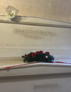
Marie Curie buried with her husband, Pierre Curie


References:

Thompson, M. K., Poortmans, P., Chalmers, A. J., Faivre-Finn, C., Hall, E., Huddart, R. A., Lievens, Y., Sebag-Montefiore, D., & Coles, C. E. (2018). Practice-changing radiation therapy trials for the treatment of cancer: Where are we 150 years after the birth of Marie Curie? British Journal of Cancer, 119(4), 389–407. https://doi.org/10.1038/s41416-018-0201-z


A Monument to Disease & Disgust

By Duke McDaniels

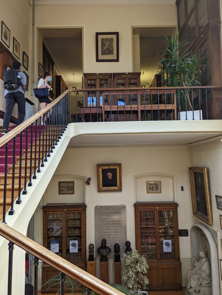
The entry stairway to the Musée des Moulages, filled with memorabilia of famous individuals integral to the development of the medical field. Photobombed by some of my classmates.

After an incredibly drama-suffused tale of miscommunications and scheduling mishaps, we of the NBB Paris Study Abroad team finally managed to make it to a visit at the Musée des Moulages, a famous dermatology museum here in the aforementioned city of Paris. While I do admit to initially rolling my eyes a bit at the prospect of visiting such a place, especially after all the trouble it had taken to get there, I wasn’t expecting the experience to be particularly valuable. How wrong I was. Visiting that monument to skincare gone-awry gave me a priceless gift I could never have gotten anywhere else in the world at my age: the 100% certainty that I would never, EVER explore the realm of dermatology as long as I lived.

The museum was particularly secretive about its contents, given that it housed wax (or perhaps plaster, I wasn’t really sure) casts and models of various dermatological diseases and disorders. The fact that these models were so unique and intricate meant that pictures weren’t allowed to be taken within the museum’s main exhibit, and when I say that every person reading this should be praising the powers that be for this rule, I mean every word. I saw things more grotesque than the minds of the already deeply-disturbed creatives within the horror genre could hope to think of on their own. With every oozing sore, swollen appendage, and fungus-eaten slab of flesh we passed, I grew evermore thankful I had skipped both breakfast and lunch that day in favor of my other homework assignments.

The more intense my feelings of revulsion at the frailty of the human form and all its vulnerabilities grew, the more I became interested in the actual source of these feelings. Disgust is an emotion commonly thought to be a response evolved to promote the avoidance of disease (Davey, 2011), a theory which makes sense given the environment which was evoking that emotion. Evidence for this evolutionary basis includes the cross-cultural facial expression associated with the emotion (i.e. wrinkled nose & downturned mouth), feelings of nausea, and avoidance of the object of disgust accompanied by a fear of being contaminated by it (Davey, 2011). The case study of myself checked all of the boxes, and in my mind, lended some more credibility to this theory. The fact that dermatologists can put themselves into such environments while thinking of nothing but aiding the afflicted person is truly admirable. It is not, however, a mindset I can share for the moment.

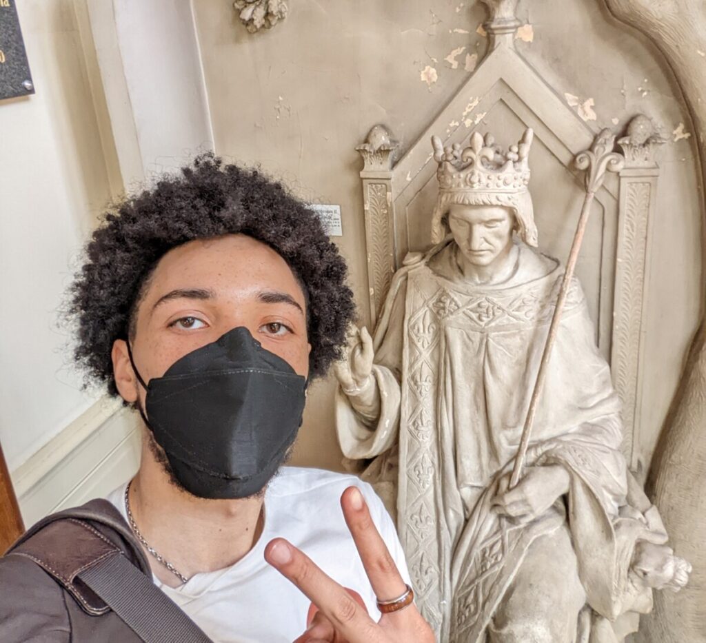
One of the few pictures I was able to take in the building. My good buddy and I, Louis IX admiring the lobby of the building, still blissfully unaware of the horrors awaiting within.

Davey, G. C. (2011). Disgust: The disease-avoidance emotion and its dysfunctions. Philosophical Transactions of the Royal Society B: Biological Sciences, 366(1583), 3453–3465. https://doi.org/10.1098/rstb.2011.0039

A Snapshot into Medical Technology

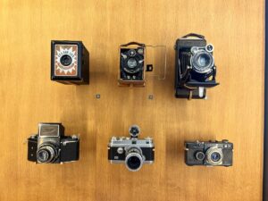
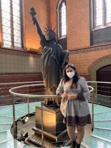
I have always loved taking photos. When preparing for any trip, the first things in my suitcase are always my cameras! My goal for Paris was to be able to document all the beautiful buildings, delicious food, and fun experiences through my camera lens. A few days ago when I first heard about our trip to the Musée des Arts et Métiers, or Museum of Arts and Crafts, on Wednesday, June 8th, I wasn’t sure what to expect. However, I was pleasantly surprised to find rows and rows of scientific instruments and inventions in front of me. The contraptions ranged from communication devices to modes of transportation and even architectural models. My favorite room as a whole was the one that housed the French replica of the Statue of Liberty, airplane models, and antique cars at the end of the museum (Figure 1). But if I had to choose one singular exhibit that called my attention the most, it was the showcase of all the different cameras throughout the years (Figure 2).

Figure 1. Here you can see the advancements of the digital camera over time.


It is fascinating to think about the major technological improvements that have been made over the past 100 years and how the invention of one item can pave an avenue of inspiration for so many other products. The development of the camera not only served as a tool for leisure and documentation but also allowed improvements in the medical field. Today, cameras are used in many procedures including neuroendoscopies. A neuroendoscopy was not a technique I was very familiar with, but it is a minimally invasive surgery tool that allows for tissue sampling in the brain usually because of brain tumors. In a retrospective descriptive study conducted by Deopujari, Shroff, Karmarkar, & Mohanty in 2022, 27 previous procedures were analyzed that utilized either endoscopic tumor biopsy (ETB) or endoscopic third ventriculostomy (ETV) techniques to treat pediatric patients. Children were laid in an upright position with their heads turned to a 20-degree angle to the left such that the right side was exposed for these procedures. Researchers were able to insert the endoscope and perform the method with just a 4-6 cm coronal suture. The study showed that the accuracy of these procedures in children with pineal region tumors “has been above 75%”, which is a statistically significant value. Neuroendoscopic biopsy as a technique is becoming much more common in the medical field as it has proven itself as a safe and effective procedure with minimal invasiveness. I can expect this technology to expand even further in the next few years as it becomes more and more effective.

Figure 2. Me posing by a French replica of the Statue of Liberty!


References: 

Deopujari, C., Shroff, K., Karmarkar, V., & Mohanty, C. (2022). Neuroendoscopy in the management of pineal region tumours in children. Child’s Nervous System. https://doi.org/10.1007/s00381-022-05561-0