A COVID-19 Memoir: “Prison” with a view

This week I spent most of my time in Hell! As many of you know, I contracted COVID-19 last week and had to quarantine for 5 days. My experience was awful. There is no sugar coating it. The virus drained me both physically and mentally.

Prior to quarantining, we went to the Soccer game which was amazing but I’m pretty sure that that’s the day of first exposure resulting in me contracting Covid.
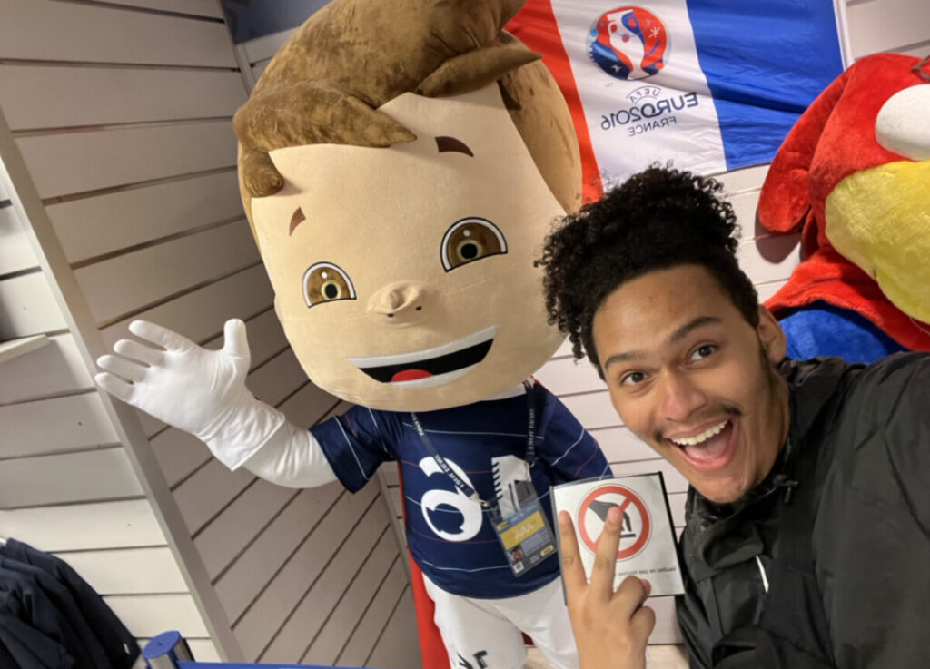
Here is me in a state of blissful ignorance prior to getting my positive result.

Fast forward to this past Monday night when I started to feel low energy. I knew that my immune system was on alert but I thought it was just a sinus problem. The next day, however, I started to feel some serious fatigued and was told to get tested. When I got the positive result, I was heartbroken. This was my first time contracting COVID so I didn’t know what to expect.

The physical symptoms were hard but manageable but the mental ones really hit me. During that time, I had felt isolated, depressed, and lonely but also simultaneously stressed about missing all this work. In thinking about this loneliness I searched for any additional information. From what I found I learned that the “lonely areas” in the brain are the amygdala as it is the emotion-center of the brain but also the Nucleus Accumbens which provides a positive reward aspect during social feedback (Lieberz et al. 2021). I also learned the NAcc is less activated during times of loneliness which may contribute to an overall lack of motivation( Lieberz et al. 2021). With no social feedback and the same environmental stimulus, no wonder why I lacked the motivation to do anything. I think it was the loneliness that was preventing me from working


One thing that helped me through all this loneliness was all the people supporting me I’m extremely grateful for my classmates who reached out and checked in on me and sent wishes to get better. These very kind messages did wonders for my immune system as well as my overall hope of getting through this. I’m even more grateful to my roommate Adway for making me soups for the past couple of days and Duke for disinfecting any surface that I touched to ensure that I would spread this horrid virus to anyone else. I’m also really grateful for my Mom and partner who were rooting for me through this dark time. Also, shout out to the two Squishamallows that my partner packed for me. These guys are the real pillars of my mental health!!
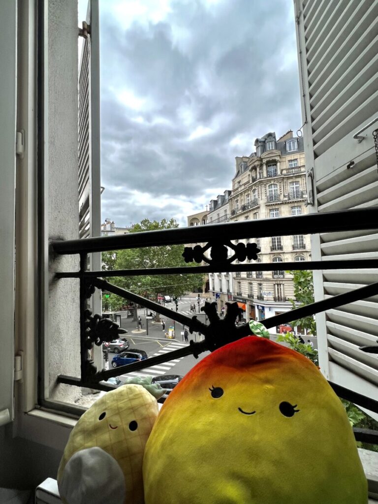
Here are my support fruits that got me through it! Left is Maui and right is Ximena. Also, the cool view where I spent most of my time people-watching can be seen in the background.

Lieberz, J., Shamay-Tsoory, S. G., Saporta, N., Kanterman, A., Gorni, J., Esser, T., Kuskova, E., Schultz, J., Hurlemann, R., & Scheele, D. (2021). Behavioral and neural dissociation of social anxiety and loneliness. https://doi.org/10.1101/2021.08.25.21262544 
Rugby (and European Football) and CTE

by Samantha Feingold

“Does anyone know what Rugby actually is?” This question floated around in several variations the week before we were to attend a France vs. Denmark rugby match.

“What?” asked our waiter several times before it was evident we weren’t pronouncing it well enough for him to understand. Lauren gestured throwing a rugby and he finally understood, but said he didn’t know the sport well enough to explain. While the restaurant is nearby our apartment and we have eaten there enough times for this waiter to recognize us when we walk past, an Italian restaurant maybe wasn’t the best place to ask about rugby.

“It’s like football but without helmets,” said a peer. It is important I emphasize at this time she was referring to American football. This statement left me more confused as helmets and padding seem to be the most important part of football, a high contact sport. Research on retired football players with chronic traumatic encephalopathy (CTE) has amplified in conjunction with the increase of media coverage on cases of severe cognitive deterioration and changes in mood in famous players (Buckland et al., 2019). However, there are minimal studies on CTE and long-term negative effects on the brain for rugby players (Buckland et al., 2019).

Prior to the match, my peers presented a crash-course on rugby and an in-depth journal article review on rugby and CTE (Zimmerman et al., 2021). It quickly became evident that rugby is quite intense, and it was likely there would be many head collisions.

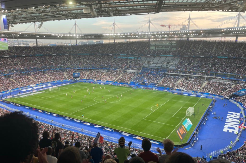
When we finally arrived at Stade de France, all the fans were bouncing with excitement and kids ran around with French-flag face paint. It wasn’t until we settled in our seats and the pre-match rituals concluded that we realized the peculiar goals and that what in front of us resembled European football far more than having any parallels to American football (Figure 1).

Figure 1. The European football field at Stade de France for the Denmark vs. France match.

While we did not see rugby that evening, we were lucky enough to watch a qualifying match for the World Cup (Figure 2)! Within minutes of realizing this was soccer and not rugby, it became evident that instead of being tackled, there was risk for brain injury from repeatedly heading the soccer ball. The potential force exerted from heading ranges from 500-1200 Newtons (Teymouri et al., 2012) and is equivalent to a maximum of over 250 pounds-force (lbf). While this is not quite the impact of a moving 300+ pound American football player, the European football players are (like the elite rugby players) not wearing helmets. Thus, while soccer is very entertaining, it is important to properly teach young players how to head a soccer ball and be aware of the possible injury.


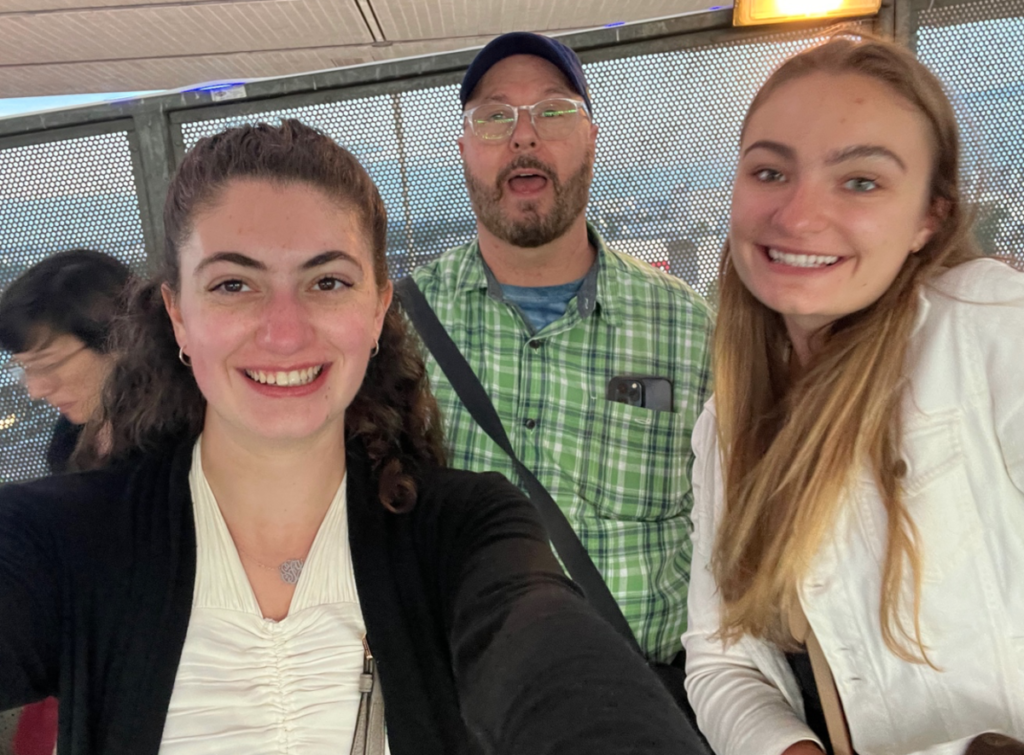
Figure 2. Samantha Feingold, Lauren Cobitz, and Dr. Easterling photographed at the Denmark vs. France soccer match.

Works Cited

Buckland, M.E., Sy, J., Szentmariay, I. (2019). Chronic traumatic encephalopathy in two former Australian National Rugby League players. acta neuropathol commun 7, 97. https://doi.org/10.1186/s40478-019-0751-1

Teymouri, M., Sadeghi, H., Nabaei, A., & Kasaeian, A. (2012). The relationship between
biomechanical-anthropometrical parameters and the force exerted on the head when
heading free kicks in soccer. Archives of trauma research, 1(1), 44–48. https://doi.org/10.5812/atr.5307

Zimmerman, K. A., Laverse, E., Samra, R., Yanez Lopez, M., Jolly, A. E., Bourke, N. J.,
Graham, N., Patel, M. C., Hardy, J., Kemp, S., Morris, H. R., & Sharp, D. J. (2021).
White matter abnormalities in active elite adult rugby players. Brain
communications, 3(3), fcab133. https://doi.org/10.1093/braincomms/fcab133

La Trephine

The rapidity at which the field of neurosurgery has continued to evolve became evident to me during this week’s visit to Musee d’Histoire de la Medicine (Museum of the History of Medicine). We began the tour by observing what, at first, seemed to be ancient hunting tools from the San people of Southern Africa. They were mismatched in size, had asymmetrical designs, and had a mix of smooth and jagged edges. I soon found out that I was staring at the first neurosurgical toolkit used by an early French neurosurgeon. It amazed me at how these rudimentary tools were used to penetrate and tinker with an organ as complex and delicate as the human brain. Even more incredible was how these tools served as the foundation for numerous generations worth of advances in neurosurgical equipment. As we progressed down the glass boxes encasing the toolkits, I noticed them to start to garner a more sophisticated form. They became increasingly symmetrical and gradually adopted a more streamlined appearance. Interestingly, I noticed how aesthetics became more of a priority. Near the beginning of the tour, prosthetic limbs had the appearance of a robotic arm. However, closer to the 19th and 20th century, the prosthetic limb, once decked out in rusted metal, was replaced with a pale tone and easily distinguishable fingers.

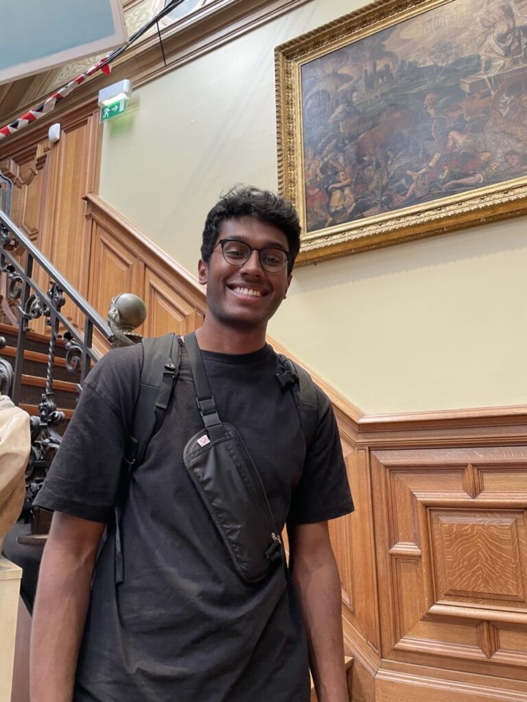
Image 1: Me at soaking in the rich medical history at Musee d’Histoire de la Medicine. 

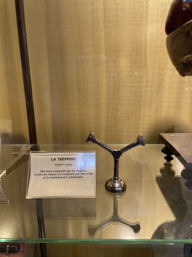
Later in the tour, I stumbled across a tool called “La Trephine” meaning “The Trepan”. In medical practice, a trepan is used to create a hole in the skull in order to expose different parts of the brain for operation. Nowadays, with more advanced technology being accessible, a cranial drill is used and is often battery-powered. The trepan in the museum, however, did not look like modern day cranial drills. It was shaped like a “Y” and had a cylindrical bottom where it attached to the brain. In order to use this tool, the surgeon would grab the top parts and twist repeatedly to make circular incisions into the skull until the bone could be removed. I discovered that this tool was commonly used to treat individuals with epilepsy. Past neuroscientists thought that mental disorders could be treated by creating an opening in the skull to allow for the demons to escape. Today, we know that epilepsy is treated with anti-epileptic drugs (AEDs) that decrease membrane excitability by interacting with neurotransmitter receptors and ion channels (Macdonald et al., 1995). This visit was very exciting and allowed me to develop a newfound understanding and appreciation for how far medicine has truly come.

Reference:

Macdonald, R. L., & Kelly, K. M. (1995). Antiepileptic drug mechanisms of action. Epilepsia, 36(s2). https://doi.org/10.1111/j.1528-1157.1995.tb05996.x

Visiting the Mona Lisa

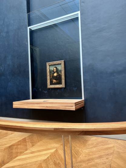
On Saturday, June 4th, myself and some other students visited the Louvre. Before coming to Paris, there were a few places I heard of or was told I had to see. The Louvre was one of them. Knowing that I was going to be able to see very few exhibits in my few hours there, the Mona Lisa was a must-see for me.

A photo I took of the Mona Lisa close enough to see the glass that saved her from the recent cake incident!  

There is quite a bit of literature explaining that art has positive impacts on mental health. While I can definitely see how this would be true, I found the museum overwhelming and anxiety-inducing due to the crowds. Maybe the word has gotten out about the neurological benefits of art and people can’t wait to go! Or maybe it was just a busy Saturday. I’ll let you decide which explanation to believe.

Christianity, COVID, and Cathedrals

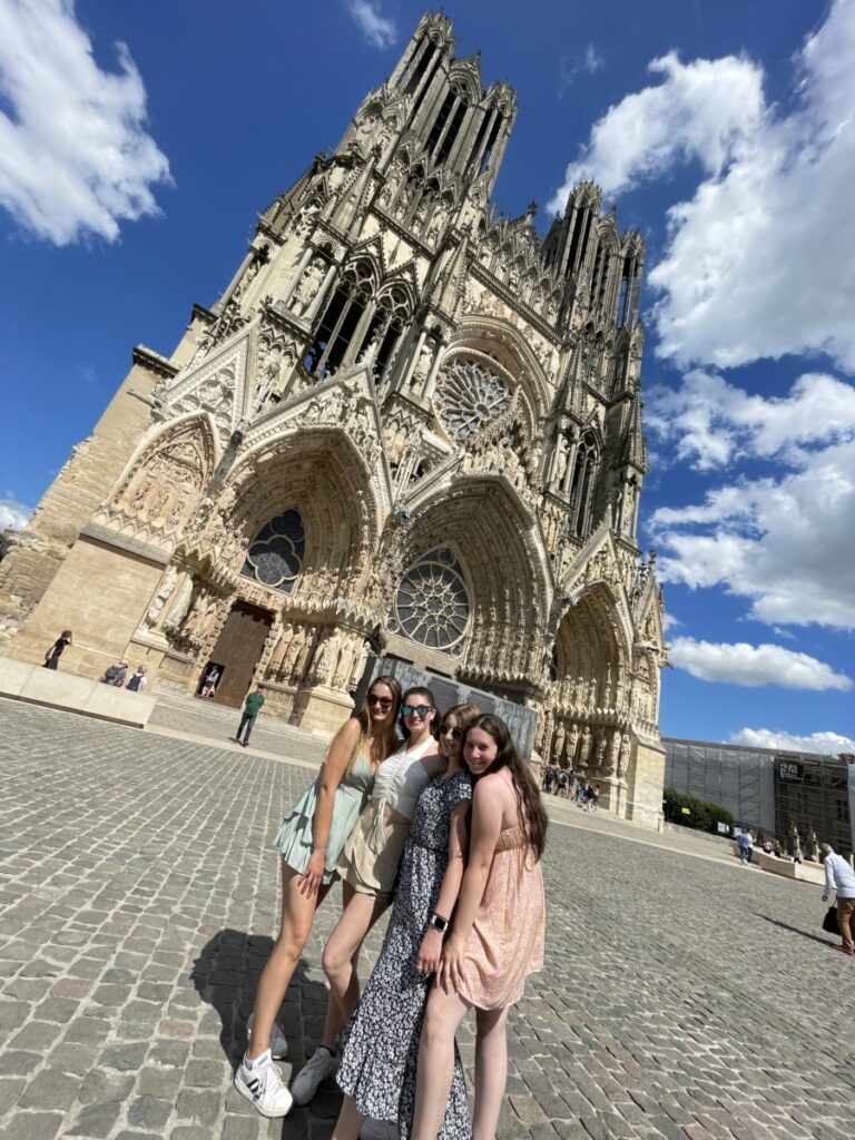
On June 11th, 2022, Sam, Rachel, Cynthia, and I visited Reims, France with Aman, one of my friends from Emory. During our visit, we saw the Notre-Dame Cathedral of Reims, which translates to “Our Lady of Reims.” This beautiful Roman Catholic cathedral, dedicated to the Virgin Mary, was the traditional location for the coronation of the kings of France. The cathedral was founded in the 5th century and has gone through numerous restorations throughout the past few centuries, including after being damaged in the French Revolution and in World War I, when it was struck by more than three hundred artillery shells. I was not only awed by beautiful building but also the deep-rooted history of Christianity in France. Christianity is as much embedded France’s history as it is ingrained in the culture and everyday lives of French citizens. For example, I found it interesting to learn that the grocery store near our apartment does not sell alcohol on Sundays.

Figure 1. A photo of Sam, Cynthia, Rachel, and I in front of the Notre Dame Cathedral of Reims

While visiting the cathedral, I reflected on the articles we discussed in NBB 402W about COVID-19, which sparked my interest in learning about the connection between religion and the pandemic since religious services quickly became hotspots for the virus. I wondered whether the illness made people feel more connected to their religious practices, so I did some research regarding the pandemic and religion. One Polish study found that most citizens surveyed declared faith/spiritually was important to them, but their faith did not strengthen in the face of the pandemic. They found that a higher percentage of Catholic respondents believed in the protective power of faith compared to other denominations and more Catholic respondents also declared that faith had a crucial role in their life (Kowalczyk et al., 2020). Despite this study showing that the pandemic did not increase spiritual belief, religion is obviously an important part of the European lifestyle. Studies have shown neurobiological changes correlated with religion and spirituality specifically in medial frontal cortex, orbitofrontal cortex, precuneus, posterior cingulate cortex, default mode network, and caudate (Rim et al., 2019), which could explain the importance of religion in people’s lives.

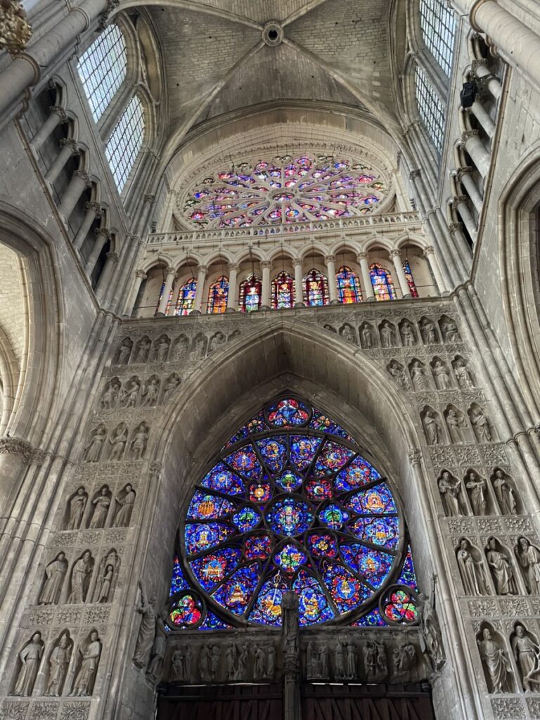
Figure 2. Picture of the stain glass windows on the inside of the cathedral

Although the outcomes of the pandemic study were not what I was expecting, I found it interesting to learn more about Christianity and Catholicism in Europe, as well as the brain changes association with religion. Overall, this visit allowed me to feel more connected to my late grandparents on my dad’s side, who were raised Catholic.

Citations:

Kowalczyk O, Roszkowski K, Montane X, Pawliszak W, Tylkowski B, Bajek A. Religion and Faith Perception in a Pandemic of COVID-19. J Relig Health. 2020;59(6):2671-2677. doi:10.1007/s10943-020-01088-3

Rim, J. I., Ojeda, J. C., Svob, C., Kayser, J., Drews, E., Kim, Y., Tenke, C. E., Skipper, J., & Weissman, M. M. (2019). Current Understanding of Religion, Spirituality, and Their Neurobiological Correlates. Harvard review of psychiatry27(5), 303–316. https://doi.org/10.1097/HRP.0000000000000232

Eiffel Tower – Work of Art or Eyesore?

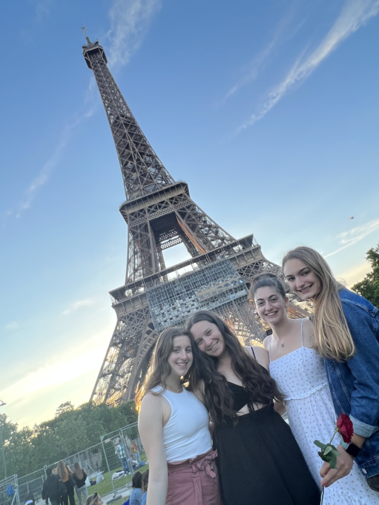
On Wednesday, June 1st, a group of students took advantage of having no major assignments due the next day and spent the evening at the Eiffel Tower. All walking towards the tower from different directions due to the locations of our various metro stops, we met under the center of the tower which we quickly learned was not the ideal location. We relocated to the lawn where we found an open area of grass to have our picnic. I enjoyed my caramel crepe while others were feasting on bread and cheese. Little did we know that we had the best photographer among us (Jacob) who gave us a little spontaneous photoshoot gifting us the ability to remember all the fun we had that night.

Photo by Jacob of me with Cynthia, Sam, and Lauren in front of the Eiffel Tower.

On our first day in Paris we did a boat tour as a class and when we passed the Eiffel tower our guide shared some interesting history about it. The tower’s construction was complete in 1889 in time for the World’s Fair and was not meant to be a permanent structure. When it was first built, Parisians were quite critical of the tower claiming that it was an eyesore and did not fit in with the city. However, despite this opinion of the locals, the Eiffel Tower has become one of the most famous landmarks and is the staple of the city.

This got me thinking about the neuroscience behind the perception of beauty. In previous classes, I have learned about perception of beauty in faces/people and the brain regions involved in that but did not know much about how this applied to objects such as buildings or art. In searching the literature, I was introduced to the field of neuroaesthetics (Iigaya et al., 2020). Neuroaesthetics explores the neural basis for judging art, of which architecture and monuments fall under. Early studies found that the brain regions involved in viewing pleasant aesthetics were also involved in reward pathways. This suggests that looking at an aesthetically pleasing work of art elicits a similar response to other rewarding stimuli. They also explained that assigning aesthetic value is done by breaking a stimulus down into elementary features to be processed. This is a concept I am familiar with from NBB 301 when we discussed sensory processing.

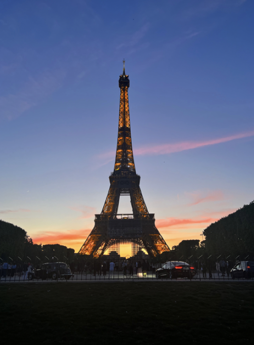
Eiffel Tower lit up as the sun is setting.

In my very unprofessional opinion, I thought the Eiffel Tower was pretty when it was lit up, but during the day it was more exciting due to its significance rather than its beauty.

Reference:

Iigaya, K., O’Doherty, J. P., & Starr, G. G. (2020). Progress and Promise in Neuroaesthetics. Neuron, 108(4), 594–596. https://doi.org/10.1016/j.neuron.2020.10.022

Faces of the Louvre


No trip to Paris would be complete without a visit to Museé de Louvre. The world’s most visited museum is housed in the beautiful 13th century Louvre Palace. It was fascinating to appreciate art in a space that was so illustrious, with the ceilings vying for equal attention to the paintings on the walls. Despite visiting on the day that the Mona Lisa was smeared with cake, my friend and I unfortunately did not bear witness to the protest (nor were we involved, no worries). We remained in the museum until closing, and still felt that we only scratched the surface of all there was to explore.

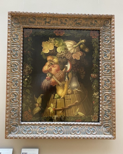
On our way out, four particular paintings caught my attention. Known collectively as The Four Seasons, these works were produced in the 14th century by Giuseppe Arcimboldo. Each one depicts a portrait composed of fruit, vegetables, and plants that relate to the respective season. Autumn represents a man but his neck is made up of pears, his chin is a pomegranate, and his ears are mushrooms. I stood there wondering why it was so familiar, and then I remembered we learned about object recognition in NBB302. Propagnosia, also known as facial blindness, is an impairment in the visual recognition of faces. Normally, faces are processed holistically but lesions in the occipital region in the ventral pathway (known as the fusiform face area) causes face blindness (Haeger et al, 2021).

Figure 1: Arcimboldo’s Autumn on display at the Louvre.

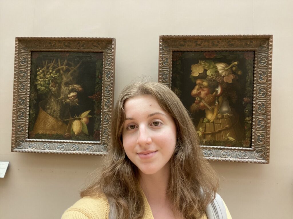
Arcimboldo’s paintings have actually been used in previous studies to observe this phenomenon. In a 2011 study, these paintings were shown to individuals with propagnosia (Rossion et al, 2011). To an unimpaired human, the elements of the painting can collectively be viewed as a face due to the object configuration. However, those with deficits in the FFA are able to only recognize the discrete components of the painting, in this case the individual fruits and vegetables. It was interesting to make this connection and it helped me more deeply understand the studies we were talking about by being able to view the stimuli in person myself.

Figure 2: Me in front of the paintings.

References:

Haeger, A., Pouzat, C., Luecken, V., N’diaye, K., Elger, C., Kennerknecht, I., … & Dinkelacker, V. (2021). Face Processing in Developmental Prosopagnosia: Altered Neural Representations in the Fusiform Face Area. Frontiers in behavioral neuroscience15.

Rossion, B., Dricot, L., Goebel, R., & Busigny, T. (2011). Holistic face categorization in higher order visual areas of the normal and prosopagnosic brain: toward a non-hierarchical view of face perception. Frontiers in human neuroscience4, 225.

Brain de Fontainebleu 

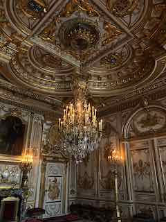
As we toured the seemingly endless halls of Chateau Fontainebleu, I thought about how distinct each room in the manor appeared. There were countless styles to admire from the gorgeous Gobelin tapestries to the baroque frescos to Marie Antoinette’s lavish furniture possession; I was overwhelmed and overstimulated. I was trying hard to connect the dots, to make sense of the rise and fall of these great epochs in French history. I felt that each corridor was a synapse between the many neuron chambers–each complete with a world of organelles from lost ages. I couldn’t make sense of it by the units, but the Chateau made sense as a composite. Perhaps this is how we must view our brains. Consciousness isn’t something so much to be understood as it is to be experienced. As we navigate our memories that construct the rooms within our minds, it should not be so much of a priority to rationalize which doors lead where (for surely, you will get lost), but rather, an exercise to clean and maintain what parts of yourself to which you are still able to access.

Pere Proust

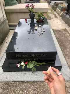
This is the grave of Marcel Proust from our class visit to the Père Lachaise Cemetery. I first discovered who he was from a reference to his work, “In Search of Lost Time.” I have not directly read his work yet, but I read Alain de Botton’s ‘self-help’ book titled, “How Proust Can Change Your Life” where I was encouraged to question many of the social norms and ways of life that most of us find ourselves stuck in. In relation to the class, many have said that Proust was actually a pioneer in neuroscience as a theorist, and modern work simply builds upon the artists of past generations. Proust shined a light upon the nature of the mind and on consciousness.

More here if you are interested:

https://www.google.com/url?sa=t&rct=j&q=&esrc=s&source=web&cd=&ved=2ahUKEwjeurnXxZH4AhVRXRoKHXFDCPIQFnoECBMQAQ&url=https%3A%2F%2Fwww.amazon.com%2FProust-Was-Neuroscientist-Jonah-Lehrer%2Fdp%2F0547085907&usg=AOvVaw2G2jG5V1EXra-FdRWTtWfu

Ballet: A Workout for Brain and Body

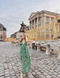
Me in my best attempt at a Versailles-esque outfit standing outside the gates to the palace after the ballet performance.

The last time I was in Paris, it was for ballet class when I was in the eighth grade. Through the language barrier and intimidation factor of taking a class in a new country, I loved my ballet studio here and the opportunity to connect with girls I couldn’t directly communicate with through an art form we all loved. So, this time when I touched down in Paris, I knew that I wanted to find a way to appreciate the thing that brought me here in the first place. After researching with my roommates, we discovered that we would be in Paris over the 250th anniversary of Marie Antoinette’s wedding and that it was being celebrated with a ballet at the Versailles Royal Opera House. So, on a rainy weekend day, we put on our most Versailles-appropriate dresses and made the hour and a half-long journey to the Opera House. 


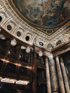
The inside of the Opera House before the show; this is the same Opera House that Marie Antoinette watched performances in while she lived at Versailles during her husband’s rule.

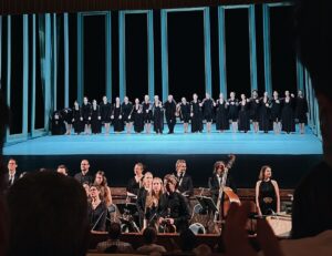
The ballet itself was incredible, with historical context provided on a screen above the stage and the dancers carefully telling the story of Marie Antoinette’s life, death, and antics on stage. Amidst the colorful costumes, contemporary take on classical ballet, and endless violin swells, I felt reconnected to my love for dance; the opportunity to experience that same passion in such a unique way was beyond incredible, and I know that the memory of that night is one I’ll cherish forever. 

Connecting this experience back to neuroscience, though, was equally as interesting. Having grown up dancing my whole life, I knew there were lessons I’d learned and habits I’d built that stick with me even today (I still find myself measuring eight-counts in music and counting steps as I walk across a room). But, in light of the courses we’re taking here, I thought it would be interesting to explore the connection between dance and the brain. Through my research, I found an interesting study which explored the relationship between dance and neural plasticity. Specifically, researchers observed structural changes in the hippocampal and grey-matter volume of dancers, as well as heightened levels of white-matter function. These physical differences then led to functional improvements in memory, body balance, and several other realms. The researchers thus concluded that dance can, in fact, improve neuroplasticity by integrating brain regions (Teixera-Machado et al, 2019). 

The dancers and orchestra taking their final bows after the performance.

Overall, I think that the opportunity to connect the purposes behind both my visits to Paris was so special, and I’m glad that I had the opportunity to find a way to bring my time here full circle. I know that being here has confirmed my love for both neuroscience and dance, and I am already researching dance studios at home to continue exploring my passions from an ever-increasingly interdisciplinary perspective. 


References:

​​Teixeira-Machado, L., Arida, R. M., & de Jesus Mari, J. (2019). Dance for neuroplasticity: A descriptive systematic review. Neuroscience and biobehavioral reviews, 96, 232–240. https://doi.org/10.1016/j.neubiorev.2018.12.010