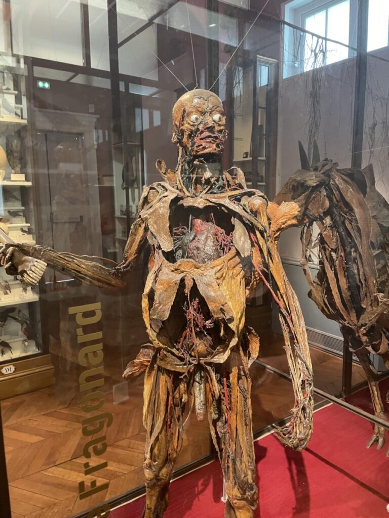
This disturbing, yet fascinating picture was taken during our class visit to the Fragonard Museum. Honore Fragonard was a French anatomist who conducted experiments that pushed ethical boundaries such as dissecting real humans and displaying them in unique action poses. From a neuroscience perspective, it was incredible to see the diffusion of veins and arteries across the body, intertwined with the delicate nerves running through the skull and down the arms. Interestingly, Fragonard also injected the veins with blue dye and the arteries with red dye, potentially also paving the way for how these organs are universally depicted in textbooks and in the classroom.
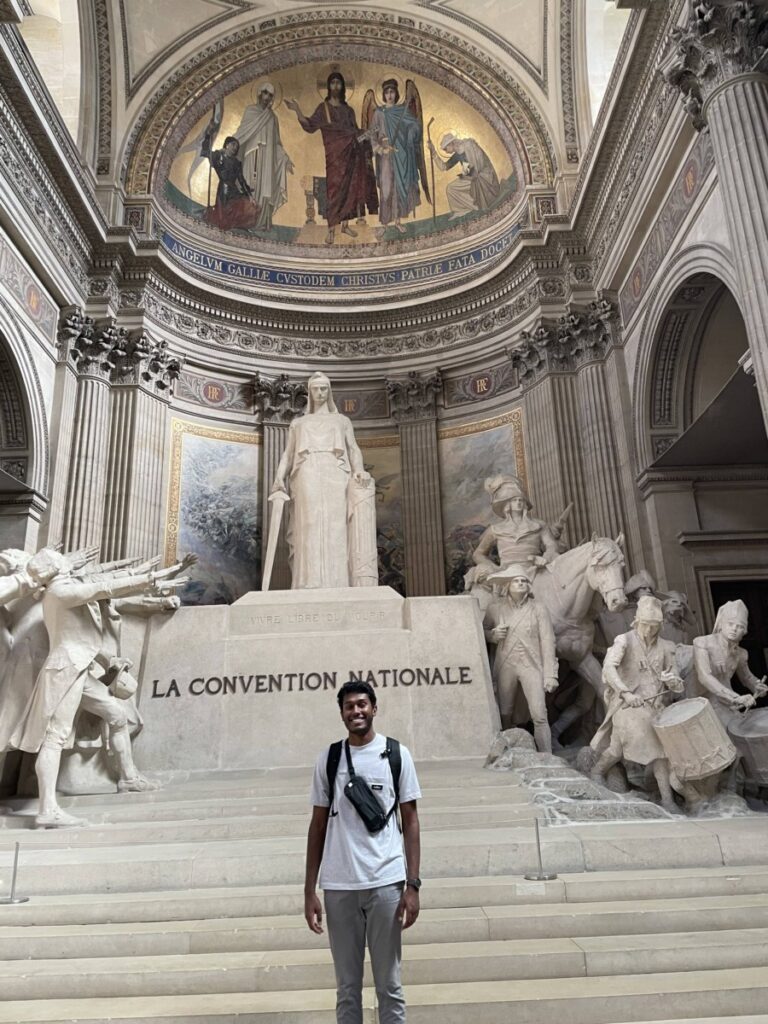
Our trip to the Pantheon was nothing short of breathtaking. The once church is now a civic building dedicated to honoring the lives of some of the greatest French citizens. One of these French greats is Marie Curie. She was a pioneering chemist who discovered the element Radium with her husband. This experience related to our NBB 402W class as her contributions have paved the way for many commonly used cancer therapeutics – such as radiotherapy for glioblastomas (Mann et al., 2018) as well as neuroimaging techniques that use radiation (sMRI, fMRI, etc.).
Earlier this week, our class took a trip to Musee de l‘Homme – an anthropological museum established in 1937. The walls leading up to the museum were decorated in brightly lit LED signs that said the word “Hello” in various different languages. It was the perfect welcome to what was going to be an afternoon filled with fun facts on the evolution of the human species.
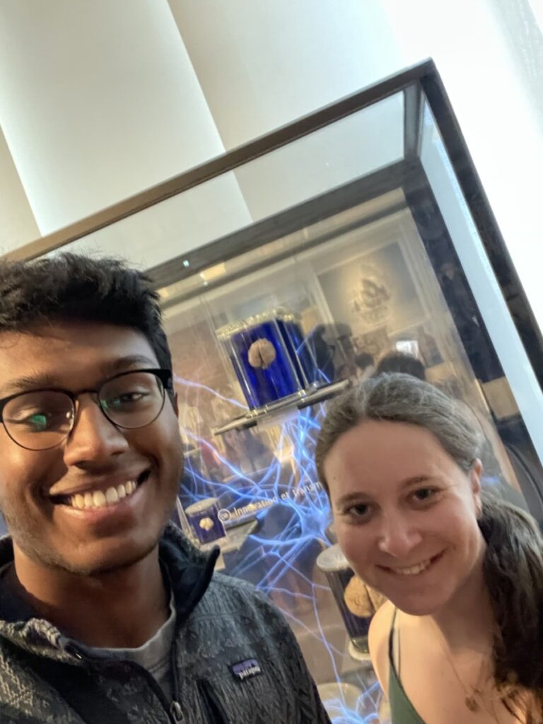
Early in the exhibit I discovered a glass case filled with the brains of different animal species such as rodents, rhesus macaques, humans, and even elephants. The brains were suspended in a hydrating solution to ensure that they did not decay. At Emory, I work in a neuroimaging lab that studies the neurostructural development of rhesus macaques brains subject to obesogenic diets and social subordination stress. Hence, it was so interesting to finally be able to see a live visual of the macaque brain, one which I have been viewing on a screen for so long. What struck me the most, however, was the drastic size difference between the human brain and the macaque brain. Since the rhesus macaque is such a common model organism for “stress on the brain” studies and is praised for its high translational value to human brains, I expected the brain size to be more similar to that of humans. However, it was surprisingly only one-third the size of a human brain. Nevertheless, numerous findings from macaque research have proven to be immensely valuable in the development of clinical treatments for humans suffering from depression, anxiety, and even mobile disabilities (e.g. the development of neuroprosthetics). One such study demonstrated that stimulation electrodes in the somatosensory cortex and recording electrodes in the anterior intraparietal area (AIP) of a male macaque can lead to tactile sensation to a NHP (Klaes et al., 2015). Such types of studies continue to revolutionize brain-machine-interface (BMI) technologies.
Image 1: Rachel and I standing in front of the encased rhesus macaque brain.
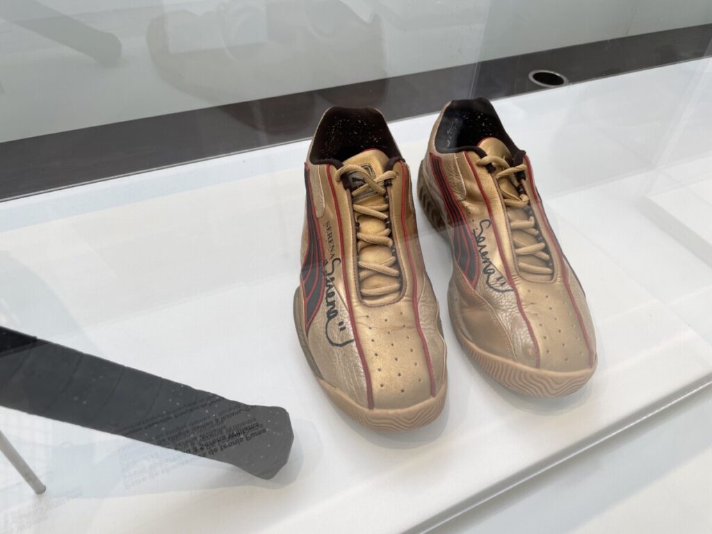
Towards the end of the exhibit, there was a section on the evolution of sneakers. As an outspoken tennis geek, I was enchanted by the display of the shoes Serena Williams wore when she won the 2002 French Open. This section was incredibly interesting as it demonstrated how cultural changes led to the evolution of various sneakers types. I have been to a sneaker museum in the US; however, this one better explained the impact of culture on sneaker evolution.
Ultimately, this trip was very eye opening for me from a neuroscience research perspective and motivated me to one day come back in the future.
Image 2: Serena Williams shoes in the sneaker exhibit that connected how human culture has shaped the evolution of sneakers.
References:
Klaes, C., Shi, Y., Kellis, S., Minxha, J., Revechkis, B., & Andersen, R. A. (2014). A cognitive neuroprosthetic that uses cortical stimulation for somatosensory feedback. Journal of Neural Engineering, 11(5), 056024. https://doi.org/10.1088/1741-2560/11/5/056024
The rapidity at which the field of neurosurgery has continued to evolve became evident to me during this week’s visit to Musee d’Histoire de la Medicine (Museum of the History of Medicine). We began the tour by observing what, at first, seemed to be ancient hunting tools from the San people of Southern Africa. They were mismatched in size, had asymmetrical designs, and had a mix of smooth and jagged edges. I soon found out that I was staring at the first neurosurgical toolkit used by an early French neurosurgeon. It amazed me at how these rudimentary tools were used to penetrate and tinker with an organ as complex and delicate as the human brain. Even more incredible was how these tools served as the foundation for numerous generations worth of advances in neurosurgical equipment. As we progressed down the glass boxes encasing the toolkits, I noticed them to start to garner a more sophisticated form. They became increasingly symmetrical and gradually adopted a more streamlined appearance. Interestingly, I noticed how aesthetics became more of a priority. Near the beginning of the tour, prosthetic limbs had the appearance of a robotic arm. However, closer to the 19th and 20th century, the prosthetic limb, once decked out in rusted metal, was replaced with a pale tone and easily distinguishable fingers.
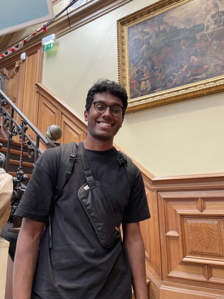
Image 1: Me at soaking in the rich medical history at Musee d’Histoire de la Medicine.
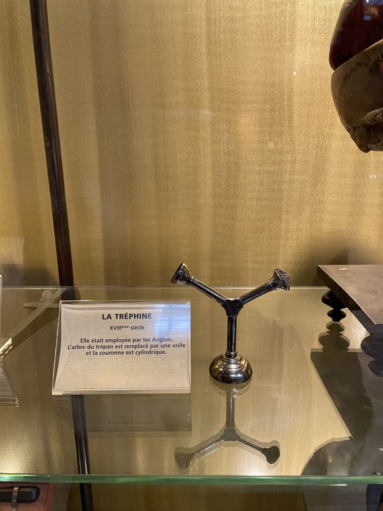
Later in the tour, I stumbled across a tool called “La Trephine” meaning “The Trepan”. In medical practice, a trepan is used to create a hole in the skull in order to expose different parts of the brain for operation. Nowadays, with more advanced technology being accessible, a cranial drill is used and is often battery-powered. The trepan in the museum, however, did not look like modern day cranial drills. It was shaped like a “Y” and had a cylindrical bottom where it attached to the brain. In order to use this tool, the surgeon would grab the top parts and twist repeatedly to make circular incisions into the skull until the bone could be removed. I discovered that this tool was commonly used to treat individuals with epilepsy. Past neuroscientists thought that mental disorders could be treated by creating an opening in the skull to allow for the demons to escape. Today, we know that epilepsy is treated with anti-epileptic drugs (AEDs) that decrease membrane excitability by interacting with neurotransmitter receptors and ion channels (Macdonald et al., 1995). This visit was very exciting and allowed me to develop a newfound understanding and appreciation for how far medicine has truly come.
Image 2. La Trephine, an instrument used in ancient neurosurgical practices to treat mental disorders.
Reference:
Macdonald, R. L., & Kelly, K. M. (1995). Antiepileptic drug mechanisms of action. Epilepsia, 36(s2). https://doi.org/10.1111/j.1528-1157.1995.tb05996.x
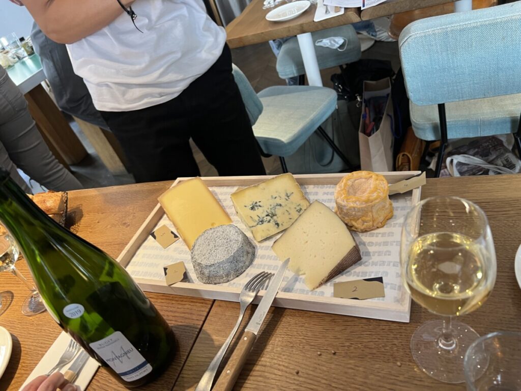
Image 2: Various types of cheese at cheese tasting.
Early in our trip, we visited Fromagerie, a cheese shop, in Paris. I have never been a huge fan of cheese; however, being able to experience the unique tastes of various cheeses native to France was memorable. In class, we discussed a paper on how cheese palatability could provide some stress relief (Fourman et al., 2021). Although cheese may not be my preferred comfort food of choice, it was nonetheless eye-opening to learn about the various cheese making techniques implemented in all parts of Europe.
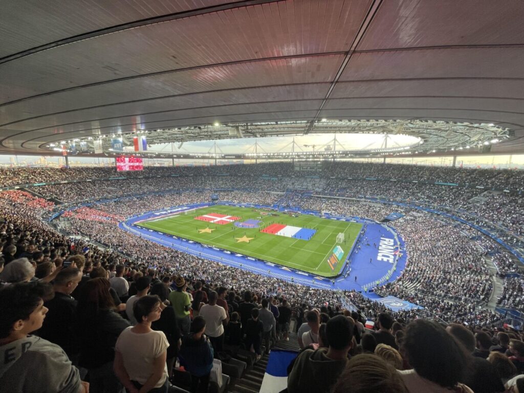
This picture was taken when our class went to a Nations League game (soccer) played between the French and Denmark national teams. The game took place at the Stade de France, undoubtedly the biggest sports stadium I had ever been to. The crowd erupted with every French possession and goal. This experience related to our NBB 402W class as we discussed how white matter tracts and brain structural changes take place with repeated blows to the head. Studies have shown that as many as 22% of soccer injuries are concussions, thus increasing the chances of neurocognitive complications for players in the future (Levy et al., 2012).
This week, our class took a trip to the Musee de Chocolat (Chocolate Museum). It did not take me long, however, to realize that our trip to Musee de Chocolat was not going to be any ordinary museum visit. Upon entering the building, we were greeted by one of the head chocolatiers. He was wearing a thick, white coat that ran to just above his ankles, and his white shoes, well, let’s just say they had seen better days. He instructed us to wash our hands, and, as the cold water trickled down my fingers… I had my EUREKA moment. We had not come here to simply learn about the history of chocolate. We were actually going to be making it!
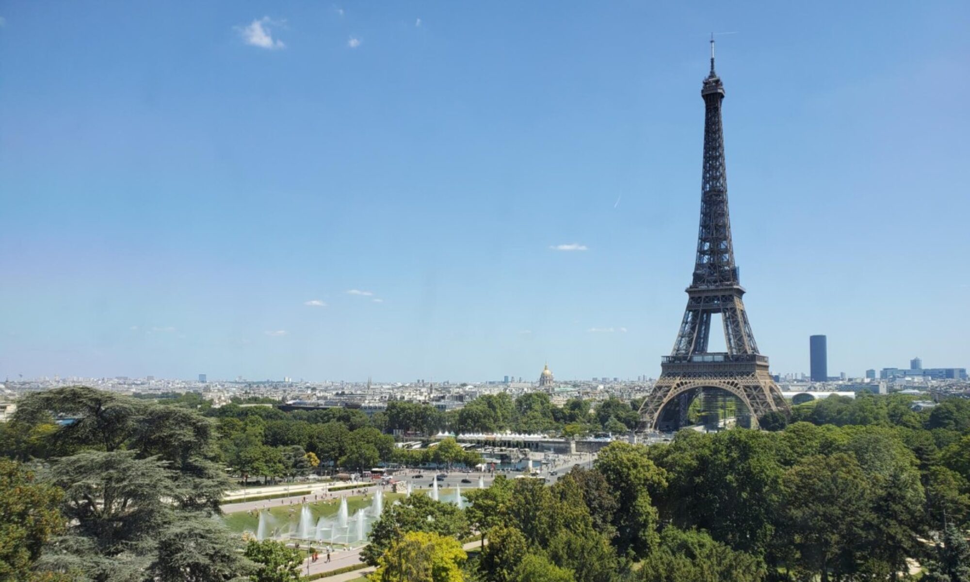
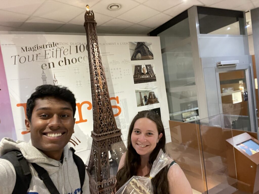
On the way downstairs, I was absolutely floored by the French architecture that the chocolatiers had brought to life with chocolate. I was most impressed by the Chocolate Eiffel Tower. It stood roughly 6 feet tall and its silky coat of brown was glistening in the light that shined from above. I asked the head chocolatier how much time it had taken to construct, and he promptly responded to me with “3 months”. Safe to say, that was just the beginning of an hour full of surprises.
Figure 1. Rachel and I in front of the Chocolate Eiffel Tower.
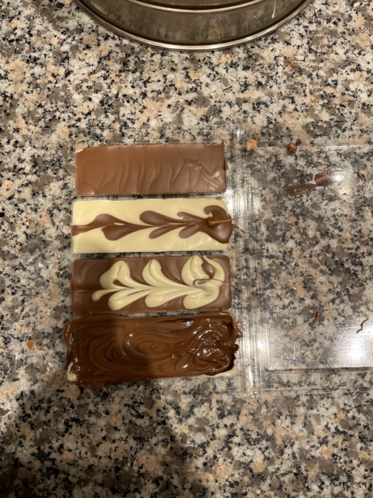
Once inside the chocolate making room, we were instructed to put on our aprons and choose a table mat to work on. Next to me were three massive, chocolate churning machines that were dripping milk chocolate, dark chocolate, and white chocolate. Our instructor conducted a few demos on how to use the chocolate to decorate our marshmallows, orange peels, and fudge blocks. He made it look incredibly easy. We strengthened our chocolate decorating skills for roughly 45 minutes then headed back upstairs to learn about the history of chocolate making and its roots stemming from Mexico.
Through this museum and my personal research, I have learned much about chocolate. In particular, I found it fascinating how brain studies have shown that dark chocolate is associated with increased verbal memory performance for two hours post consumption (Lamport et al., 2020). This is most likely due to the effects of dark chocolate having increased flavanol-rich cocoa which increases cerebral blood flow during the first 2-4 hours after first intake (Sorond et al., 2008). I was able to gather so much information from this trip with the added bonus of strengthening my chocolate making skills. Even better, I found a reason to continue indulging in this creamy treat, within reason, of course!
Figure 2. My poor attempt at making chocolate bars.
References:
Lamport, Daniel J., et al. “Beneficial Effects of Dark Chocolate for Episodic Memory in Healthy Young Adults: A Parallel-Groups Acute Intervention with a White Chocolate Control.” Nutrients, vol. 12, no. 2, Feb. 2020, p. 483. PubMed Central, https://doi.org/10.3390/nu12020483.
Sorond, Farzaneh A., et al. “Cerebral Blood Flow Response to Flavanol-Rich Cocoa in Healthy Elderly Humans.” Neuropsychiatric Disease and Treatment, vol. 4, no. 2, Apr. 2008, pp. 433–40. PubMed Central, https://www.ncbi.nlm.nih.gov/pmc/articles/PMC2518374/.
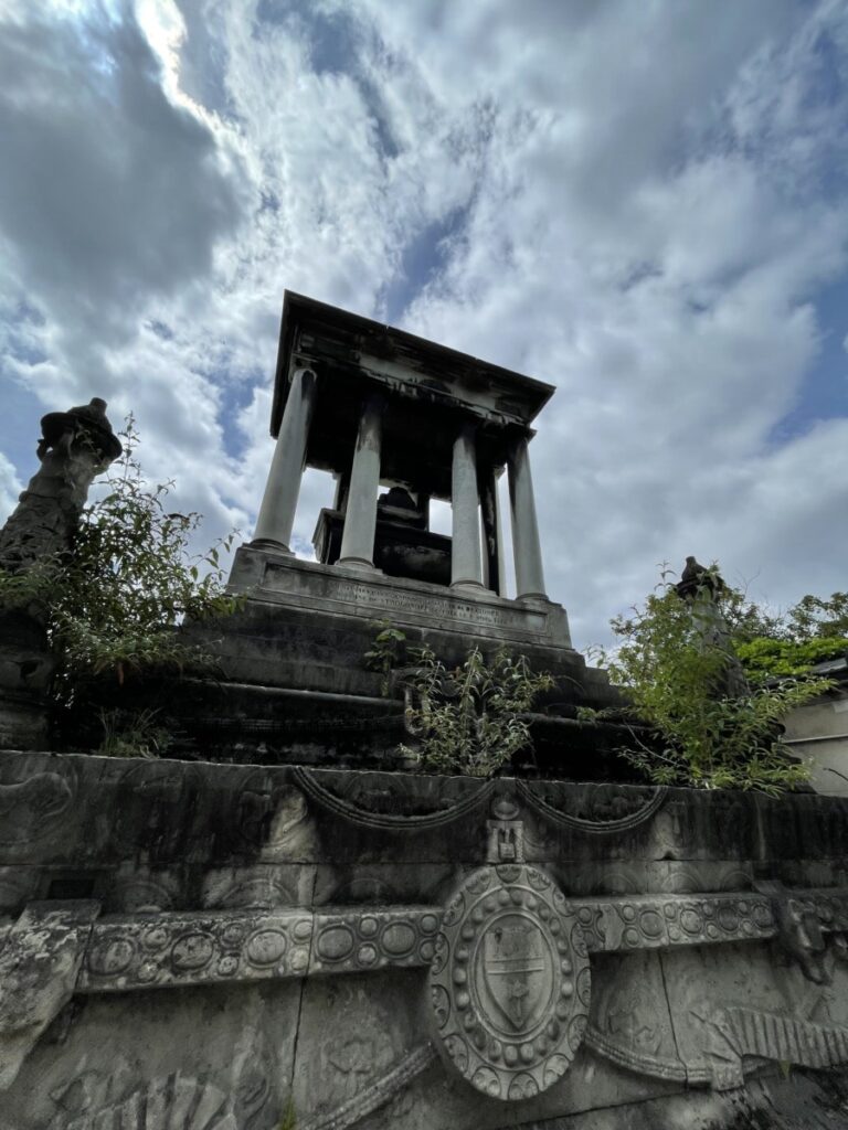
This week I had the chance to visit the Cimetiere du Pere Lachaise – arguably one of the world’s most famous cemeteries. Immediately upon entering the grounds, I was astonished by the intricate details that lined each of the tombs. From the gently carved Greek mythological figures to the gleaming crosses that could be spotted from miles away, each tomb was decked in masterful art that paid tribute to the life-long work of various well-established individuals. In the US, I have visited Arlington National Cemetery; however, my experience there couldn’t have been more different from my experience at Cimetiere du Pere Lachaise. At Arlington, all of the gravestones, similar in size and decor, were tributes to those who received the Medal of Honor. Contrastingly, the tombs at Cimetiere du Pere Lachaise were not only more majestic in nature – potentially due to the French’s appreciation for high value artwork – but also there was a greater diversity in the types of individuals honored. Prior to this visit, I had expected most of the tombs to be tributes to French philosophers, priests, physicians, and more. However, I was pleasantly surprised when I stumbled across the tomb of Jehangir Ratanji Dadabhai (JRD) Tata – an industrial entrepreneur of French and Indian descent whose work sparked the genesis of one of the biggest automobile industries “Tata”.
Figure 1: Intricately designed tomb massive in size.
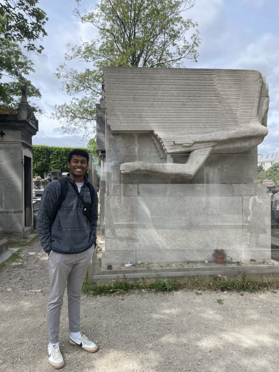
One of the tombs that stood out to me the most was that of Irish poet and novelist, Oscar Wilde. Although famous for his novels, Oscar Wilde was also interested in physiology. He wrote at a time when neuroscience threatened the status quo – that being humans have free will. Wilde, however, went against the grain and was a stout determinist himself. He rejected the notion that all humans were autonomous beings and self-determining. In fact, Wilde’s perception of free will was quite advanced for his time as recent studies have shown that the activity of certain neurons can predict behaviors before they happen. A 2018 study by Passecker et al. demonstrated that there are specialized neurons in the prelimbic cortex of rats that predict the next choice behavior during a gambling task. These findings have interesting implications as it shows the potential inability for living organisms to not have a choice in their actions because they are restricted by the electrophysiology of their neurons.
This trip helped me understand the grandeur of French culture while also exposing me to the diverse cultural perspectives on free will and the brain!
Figure 2: Adway in front of Oscar Wilde’s tomb.
References:
Cohn, Elisha. “Chapter 2 – Oscar Wilde and the Brain Cell.” Progress in Brain Research, edited by Anne Stiles et al., vol. 205, Elsevier, 2013, pp. 19–39. ScienceDirect, https://doi.org/10.1016/B978-0-444-63273-9.00002-2.
Passecker, Johannes, et al. “Activity of Prefrontal Neurons Predict Future Choices during Gambling.” Neuron, vol. 101, no. 1, Jan. 2019, pp. 152-164.e7. PubMed Central, https://doi.org/10.1016/j.neuron.2018.10.050.