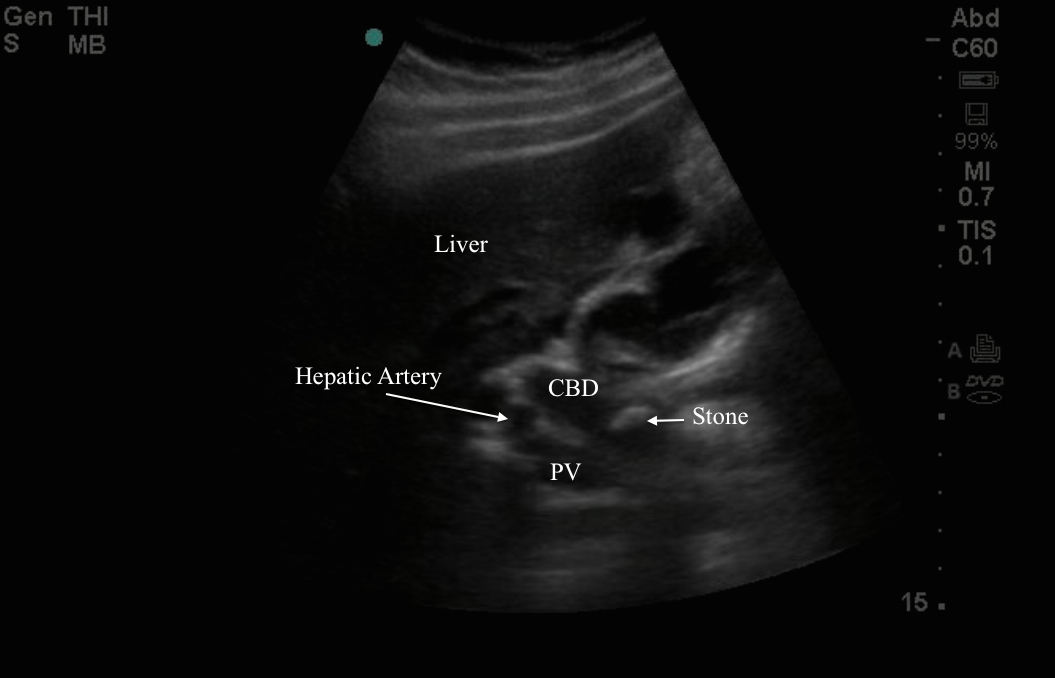
The IOW this week comes from Drs. Dustin Hill and Katie Dean. Their patient is 40’s year old male, who presented to the ER with fever, tachycardia, and RUQ abdominal pain. He had a history of cholecysttitis previously and had undergone cholecystectomy. Bedside ultrasound below was obtained. It can be challenging to get oriented when imaging patients after cholecystectomy. To begin, scan through the liver and try to identify the portal triad. Remember the portal triad contains the portal vein, hepatic artery, and common bile duct and will typically looks like “Mickey Mouse” in short axis orientation. Color flow imaging may help you to identify which structure are vascular. In the image below we see the portal triad in a long axis orientation. The CBD will run superior and parallel to the portal vein in this orientation. The hepatic artery will be sandwiched between the CBD and portal vein in a short axis orientation. After a cholecystectomy a normal CBD will measure 1cm or less. In this image the CBD is markedly dilated with sludge. On continued scanning a 1.6cm obstructing stone is seen in the CBD (image 2). LFT revealed obstructive pattern and elevated bilirubin. Diagnosis of cholangitis was made, the patient was started on antibiotics, and admitted for ERCP. Following admission sphincterotomy was performed with expression of pus, stone was extracted, and patient recovered well.
Image 1