 How do proteins fold to their functional three-dimensional structures? A random search of all of the conformational degrees of freedom available to the polypeptide chain would require a time longer than the age of the universe, and yet proteins rapidly find their native structures, sometimes within microseconds. The answer lies in the energy landscape for protein folding, shown in the cartoon at right, which is thought to have a strong energy bias towards the native structure. We seek to map this energy landscape, in order to understand how proteins fold in detail, and what happens when they misfold with disastrous consequences as in Alzheimers and other misfolding diseases.
Our approach closely integrates experiment and simulation to answer these 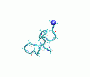 questions. We have designed experimental methods to quantitatively test the predictions of MD simulations of ultrafast folding proteins. In turn, we expect MD simulations to motivate new experiments, or help in the interpretation of experimental observables. We find such a close interplay between experiment and theory greatly benefits both, and ultimately improves our understanding of how proteins fold. Fundamentally new approaches to the rapid initiation and structure specific characterization of folding reactions are required to map the folding energy landscape. We have pioneered the development of such approaches in my laboratory, establishing the viability of laser-induced T-jump and pH-jump coupled with time-resolved spectroscopic methods. We emphasize time-resolved infrared and fluorescence techniques as well established, structure-specific probes of folding dynamics. We focus on small ultrafast folding proteins and peptide models to facilitate quantitative comparison of experiment with the predictions of folding theory and simulations.
Funding
NIH R01 GM53640-019, “Fast Events in Protein Folding,” (Dyer, P.I.), 5/2013-4/2018
Recent Publications
- “Stability of HA2 Prefusion Structure and pH-Induced Conformational Changes in the HA2 Domain of H3N2 Hemagglutinin.” M. W. Eller; H. M. H. Siaw; R. B. Dyer. Biochemistry 2021, link
- “Kinetics of Histidine-Tagged Protein Association to Nickel-Decorated Liposome Surfaces,” G. Raghunath; R.B. Dyer. Langmuir, 2019 , 35, 12550-12561 link
- “Active Site Glu165 Activation in Triosephosphate Isomerase and its Deprotonation Kinetics,” H. Deng; R.B. Dyer; R. Callender. J. Phys. Chem. B., 2019 link
- “Dynamics of hemagglutinin-mediated membrane fusion,” M.W. Eller, R.B. Dyer. PNAS, 2018 link
- “Heterogeneity in the Folding of Villin Headpiece Subdomain HP36,” S. Nagarajan, S. Xiao, D.P. Raleigh, R.B. Dyer. J. Phys. Chem. B, 2018 Just Accepted link
- “Peripheral Protein Unfolding Drives Membrane Bending,” Siaw, H.M.H.; Raghunath, G.; Dyer, R.B. Langmuir, 2018, 34, 8400–8407. link
- “Binding, folding and insertion of a β-hairpin peptide at a lipid bilayer surface: Influence of electrostatics and lipid tail packing,” Reid, K.A.; Davis, C.M.; Dyer, R.B.; Kindt, J.T. Biochimica et Biophysica Acta- Biomembranes, 2018, 3, 792- 800 link
- “Proton Transport Mechanism of M2 Proton Channel Studied by Laser-Induced pH Jump”, B. Jeong and R. B. Dyer, J. Am. Chem. Soc. 2017
- “Submillisecond Dynamics of Mastoparan X Insertion into Lipid Membranes”, E. E. Schuler, S. Nagarajan, and R. B. Dyer, J. Phys. Chem. Lett. 2016, 7 (17), 3365–3370
- “The Role of Electrostatic Interactions in Folding of β-Proteins.”, C. M. Davis, R. B. Dyer, J. Am. Chem. Soc. 2016, 138(4), 1456–1464.
- “Fast Helix Formation in the B Domain of Protein A Revealed by Site-Specific Infrared Probes.”, C. M. Davis, A. K. Cooper, and R.B. Dyer, Biochemistry, 2015, 54(9), 1758-1766.
- “Submillisecond mixing in a continuous-flow, microfluidic mixer utilizing mid-infrared hyperspectral imaging detection.”, D. P. Kise, D. Magana, M. J. Reddish, and R.B. Dyer, Lab Chip, 2014, 14, 584-591.
- “WW Domain Folding Complexity Revealed by Infrared Spectroscopy.”, C. M. Davis, and R.B. Dyer, Biochemistry, 2014, 53(34), 5476-5484.