- Evidence behind BP reduction after ICH: http://pulmccm.org/main/2013/randomized-controlled-trials/in-intracerebral-hemorrhage-rapid-blood-pressure-reductions-were-safe-interact2/
- Benzo Equivalent dosing calculator: http://www.globalrph.com/benzodiazepine_calc.htm
- Calcium Channel and Beta Blocker Toxicity review podcast (peds focused, but generalizable): http://www.pemed.org/blog/2013/4/15/calcium-channel-blockers-beta-blockers-and-undifferentiated.html
Feb 01
FOAM of the Week – Intracranial Hemorrhage, Benzo Equivalents, Calcium Channel and Beta Blocker Toxicity
Jan 26
Lit of the Week – Targeted Temperature Management (TTM)
Clinical question / background:
- In patients with ROSC following cardiac arrest, does cooling patients to 33 degrees Celsius increase the rate of survival after resuscitation compared to cooling to 36 degrees Celsius?
Design:
- Unblinded, parallel-group, randomized, controlled trial
- 939 participants in 36 European and Australian centers
- Inclusion criteria:
- Witnessed arrest
- Unconscious at presentation
- Presumed cardiac origin of arrest
- Age >18
- More than 20 minutes of ROSC after resuscitation
- Exclusion criteria:
- No ROSC within 240 minutes of presentation
- Unwitnessed arrest with initial rhythm of asystole
- Suspected or known acute intracranial hemorrhage or stroke
- Body temperature <30 degrees Celsius
Intervention:
- Reduce core temperature (bladder temp) to 33 C or 36 C after ROSC. Maintain temperature goals for 28 hours, at which point allow for passive rewarming but maintain temperature below 37.5 C for 72 hours after ROSC
Control:
- None
*Both groups sedated with for 36 hours after ROSC by providers’ sedation agents of choice.
Primary endpoint:
- Mortality at the end of the trial
Secondary endpoints:
- Favorable neurologic outcome at 180 days after cardiac arrest defined by Pittsburgh cerebral performance categories (CPC) (1 good recovery, 2 moderate disability, 3 severe disability, 4 vegetative state, 5 death) or the Modified Rankin Criteria (0 representing no symptoms, 1 no clinically significant disability, 2 slight disability, 3 moderate disability, 4 moderately severe disability, 5 severe disability, and 6 death.)
- Mortality at 180 days
Results:
- No difference in mortality at the end of the trial
- 50% vs 48% (p=0.51)
- No difference in neurologic outcome at 180 days
- CPC 3-5: 54% vs 52% (p=0.78)
- Modified Rankin 4-6: 52% vs 52% (p=0.87)
- No difference in mortality at 180 days
- 48% vs 47% (p=0.92)
Take-home:
- No difference in mortality or neurologic outcomes in patients who are cooled to 33 C vs 36 C who present with witnessed arrest of presumed cardiac cause.
Weaknesses / Critiques
- Unblinded
- No control group
- Took nearly 12 hours to cool patients in this trial, compared to 4-6 in previous trials
- Possible that smaller subset of patients (i.e. those with initial shockable rhythm) could benefit from therapeutic hypothermia
Real World Application
- Consider hospital protocol and patient’s characteristics before initiating therapeutic hypothermia
Additional information:
EMRAP October 2015 – Targeted Temperature Management 1 Year later
www.emrap.org/episode/october/october
Source:
Jan 23
FOAM of the Week – EtOH Withdrawal, Cannabinoid Hyperemesis Syndrome, Depakote Toxicity
- A couple podcasts from EMRAP covering both treatment of severe EtOH withdrawal as well out outpatient treatment of mild EtOH withdrawal:
- Phenobarb monotherapy for EtOH withdrawal: http://emcrit.org/pulmcrit/phenobarbital-monotherapy-for-alcohol-withdrawal-simplicity-and-power/
- Overview of cannabinoid hyperemesis syndrome from LITFL: http://lifeinthefastlane.com/therapeutic-showering/
- Finally, overview of valproic acid toxicity: http://www.aliem.com/valproic-acid-induced-hyperammonemic-encephalopathy/
Jan 21
Lit of the Week – Wells Score
Clinical question / background:
- Is there a simple clinical model to rule out pulmonary embolism in patients presenting to the emergency department?
Design:
- Prospective cohort study
- 930 participants in 4 Canadian Centers
- Inclusion: Adults with suspected PE with sxs < 30 days; chest pain or shortness of breath acute in onset
- Exclusion: UE DVT as likely source of PE, no sxs within 3 days of presentation, expected survival < 3 months, anticoagulation therapy for 24 hours or more, contraindication to contrast, pregnancy, < 18 y/o, unable to follow-up
Intervention:
- Implementation of clinical model to determine probability of PE
- 3 points – Clinical Signs/Sxs of DVT
- Objectively measured leg swelling and/or pain with palpation of deep veins
- 1.5 points – Tachycardia (HR> 100)
- 1.5 points – bed rest (except going to bathroom) for 72 hours, or surgery within previous 4 weeks
- 1.5 points – prior dx of DVT or PE
- 1 point – hemoptysis
- 1 point – malignancy (current rx, palliative care, or treatment within the preceding 6 months)
- 3 points – PE as most likely or as likely as alternative diagnosis based on physical exam and basic workup (EKG, CXR, screening labs)
- 3 points – Clinical Signs/Sxs of DVT
- Pretest Probability of PE based on Score
- < 2.0 points – low risk
- 2.0 – 6.0 points – moderate risk
- >6.0 points – high risk
- Primary Outcome
- 3-month occurrence of PE based on initial risk stratification
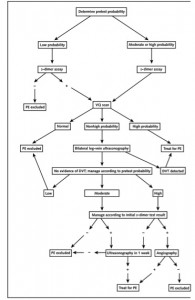
- Algorithm (see below)
Results:
- LOW pre-test probability – PE diagnosed in 1.3% at 3-month f/u
- MODERATE pre-test probability – PE diagnosed in 16.2% at 3-month f/u
- HIGH pre-test probability – PE diagnosed in 37.5% at 3-month f/u
Take-home:
- The Wells’ Criteria risk stratifies patients for pulmonary embolism (PE) and provides an estimated pre-test probability. The physician can then chose what further testing is required for diagnosing pulmonary embolism (I.E. d-dimer or CT angiogram or V/Q
Strengths:
- Simple to use; clear cut-offs
- Validated multiple times in multiple settings since original paper
Weaknesses / Critiques
- Subjective component of PE being most likely diagnosis can push score to intermediate range and lead to unnecessary testing
- Reliance on d-dimer for decision-making
Follow-up / Real World Application
- If patient determined to be low Risk, consider d-dimer testing
- Also in low-risk patients, can be used with the PERC as rule-out for PE
- The PERC rule can be applied to patients where the diagnosis of PE is being considered, but the patient is deemed low-risk. A patient deemed low-risk by physician’s gestalt who is also <50 years of age, with a pulse <100 bpm, SaO2≥ 95%, no hemoptysis, no estrogen use, no history of surgery/trauma within 4 weeks, no prior PE/DVT and no present signs of DVT can be safely ruled out and does not require further workup
- In medium / high risk patients, consider CTA (+/- d-dimer) or V/Q
- Calculator links
- http://www.mdcalc.com/wells-criteria-for-pulmonary-embolism-pe/
- http://www.mdcalc.com/perc-rule-for-pulmonary-embolism/
Jan 20
Image of the Week – Pseudoaneurysm
The image of the week comes to us from Dr Mody who used ultrasound to assess a patient with Leg Swelling two weeks after a GSW.
Jan 20
Lit of the Week – Therapeutic Hypothermia
Clinical question / background:
- In patients with ROSC following cardiac arrest due to ventricular fibrillation, does mild systemic hypothermia increase the rate of neurologic recovery after resuscitation?
Design:
- Unblinded, parallel-group, randomized, controlled trial
- 275 participants in 5 European centers
- Inclusion criteria:
- Witnessed cardiac arrest
- Initial rhythm of Vfib or non-perfusing ventricular tachycardia
- Presumed cardiac origin of arrest
- Age 18-75
- Interval of 5-15 minutes from patient collapse to first attempt at resuscitation by emergency medical personnel
- Interval of less than 60 minutes from collapse to ROSC
- Exclusion criteria:
- Temp< 30 C on admission
- Prior to arrest, comatose due to CNS depressant medication
- Pregnancy
- Response to verbal commands after ROSC
- MAP<60 for more than 30 minutes after ROSC
- Hypoxemia for more than 15 minutes after ROSC (SpO2<85%)
- Terminal illness prior to arrest
- Cardiac arrest after arrival of emergency medical personel
- Known pre-existing coagulopathy
Intervention:
- Reduce core temperature (bladder temp) to 32-34 C within four hours of ROSC. Maintain temperature goals for 24 hours, at which point allow for passive rewarming.
Control:
- Normothermia
*Both groups sedated with midazolam and fentanyl. Paralyzed with pancuronium.
Primary endpoint:
- Favorable neurologic outcome within 6 months after cardiac arrest defined as Pittsburgh cerebral performance categories 1 (good recovery), 2 (moderate disability). Poor outcome defined at category 3 (severe disability), 4 (vegetative state), 5 (death).
Secondary endpoints:
- Mortality within 6 months
- Complication rate within 7 days
Results:
- TH associated with improved neurologic outcome at 6 months: Pittsburgh cerebral performance category 1 or 2
- 55% vs 39% (p=0.009)
- TH associated with decreased rate of death at 6 months
- 41% vs 55% (p=0.02)
- No difference in rate of complications
- 73% vs 70% (p=0.09)
Take-home:
- Witnessed cardiac arrest, VF and ROSC within 1 hour should be treated with therapeutic hypothermia for 24 hours for improved neurologic outcome.
Weaknesses / Critiques
- Small trial
- Unblinded
- Only 8% of eligible patients enrolled in trial
- Follow-up study in 2013 shows no difference between 33 C and 36 C, suggesting that hypothermia not of benefit, but hyperthermia dangerous.
Real World Application
- Improved neurologic outcome: NNT of 6
- Mortality benefit: NNT 7
Further reading
- Nielsen N, et al. “Target Temperature Management 33°C vs. 36°C after Out-of Hospital Cardiac Arrest”. The New England Journal of Medicine. 2013. 369(23):2197-2206.
Jan 18
Weekly Report – Thyroid Storm
Presentation: 50 yo F with PMH of hypertension presents for shortness of breath and altered mental status. The patient is lethargic in obvious respiratory distress. Her BP is 230/130, HR is 170, RR is 35, O2 is 90% on NRB, and Temp is 38.1. She has crackles throughout all lung fields. She is intubated for respiratory failure on arrival. Postintubation a significant goiter is noted.
- Thyroid storm is a rare endocrinologic emergency that carries a high mortality if unrecognized. Symptoms are those of extreme hyperthyroidism, including hyperthermia, tachycardia, tremors, heart failure, GI symptoms, and neurologic symptoms. Patients typically have a history of hyperthyroidism (which may be previously undiagnosed) which is exacerbated by a stressor, such as infection, trauma, surgery, MI, or an iodine load (ie iodinated contrast or amiodarone).
- Diagnosis is made by clinical criteria and clinical suspicion. A high level of clinical suspicion should be held in patients with thyrotoxicosis and evidence of systemic decompensation, typically respiratory failure or significant alterations of mental status. Scoring criteria have been developed to guide diagnosis:

Recommended treatment has four components:
- Beta Blockade
- Propranolol
- 60-80 mg q4hr PO or via NG tube
- 1 mg IV q15min to effect
- Inhibits T4 to T3 conversion
- Esmolol
- Easily titratable to effect in critically ill patients
- Propranolol
- Thionamide
- Methimazole
- 60-80 mg/day PO or via NG tube
- Preferred by most endocrinologist as is less hepatotoxic than PTU
- Propylthiouracil
- 500-1000 mg load, then 250 mg q4hr PO or via NG tube
- Inhibits T4 to T3 conversion
- Preferred during pregnancy as methimazole is teratogenic
- Methimazole
- Iodine
- Saturated Solution of Potassium Iodide (SSKI) 5 drops orally q6hr
- Administer 1 hour after thionamide so as not to exacerbate storm
- Hydrocortisone
- 300 mg IV loading dose, then 100 mg q8hr IV
Case Conclusion: TSH returned undetectable while T4 and T3 were markedly elevated. She was started on methimazole, SSKI, Hydrocortisone and Esmolol GGT with improvement in vital signs. Antibiotics were given for likely pneumonia as precipitating factor. She was weaned from the vent and went home 2 weeks later with a new diagnosis of hyperthyroidism and plans for further outpatient workup and treatment.
Jan 14
Lit of the Week – ARDSNet Ventilation
Clinical question / background:
- In patients with ARDS, does ventilation with lower tidal volumes versus traditional higher tidal volumes reduce death and ventilator-free days?
Design:
- Randomized, single-blinded, controlled trial
- 861 participants in 10 U.S. centers
- Inclusion: mechanically ventilated patients with ARDS
- ARDS: Bilateral opacities on CXR/CT present within 1 week of known clinical insult not explainable by cardiogenic edema, lung effusions/nodules/collapse WITH impairment in oxygenation defined by ratio PaO2/FiO2 < 300 (FiO2 as decimal e.g. 0.21 instead of 21%)
- Exclusion: pregnancy, chronic lung disease, severe burns (>30% TBSA), patients with neuromuscular disease, < 18 y/o
Intervention:
- Low tidal volume ventilation – 6 ml/kg/breath (ideal body weight)
- Plateau pressure < 30 cm water
Control:
- Traditional tidal volume ventilation – 12ml/kg/breath (ideal body weight)
- Plateau pressure < 50 cm water
Results:
- Lower tidal volume ventilation associated with reduced mortality
- 31.0% vs 39.8% (p=0.007)
- Lower tidal volumes associated with increased ventilator-free days
- 12+/-11 days vs 10 +/-11 days (p=0.007)
- Lower tidal volumes associated with fewer days without non-pulmonary organ failure (circulatory, renal, liver, coagulative)
Take-home:
- Adult patients with acute respiratory distress syndrome should be ventilated with tidal volumes of 6 ml/kg, limiting plateau pressures to 30 cm water
Strengths:
- Well-designed, strong power to detect difference in clinical outcomes
Weaknesses / Critiques
- Single-blinded, so physicians aware of allocation, potentially biased in care provided
- Protocol allowed for varying PEEP levels to control for acidemia, may have favored intervention group + confounded
- Auto-PEEP in intervention group (due to high respiratory rate) possibly contributed to favorable oxygenation
- Addressed in post-hoc analysis and proven to be non-factor as was permissive hypercapnia in intervention group
Follow-up / Real World Application
- Foundation of tidal volume strategy in mechanical ventilation on ICU patients with ARDS
- Cited in Cochrane review — Petrucci N, Iacovelli W. Lung protective ventilation strategy for the acute respiratory distress syndrome. Cochrane Database Syst Rev. 2007 Jul 18;(3)
- ARR 10%, 28-day mortality benefit with NNT of 10
Jan 12
Image of the Week – Upper Extremity DVT
The image of the week comes to us from Drs Almebash, Liska, Sizemore, and Allen who used ultrasound to assess a patient with a history of malignancy who was found on exam to have isolated LUE Swelling.
Thanks for all of your great images this week!
Happy Scanning!
Sierra Beck MD
Jan 12
FOAM of the Week – Chemical Terrorism, NOAC Reversal, Necrotizing Soft Tissue Infections
- A timely overview on chemical, biological, radioactive and nuclear threats from St Emlyns, including info on irritant gases, nerve agents, and vessicants: http://stemlynsblog.org/cbrn-an-introduction/
- Learn about the literature behind Praxbind: http://rebelem.com/noreversaban/
- Look out for Andexanet Alfa on the horizon: http://www.emlitofnote.com/2015/11/the-great-reveal-of-andexanet-alfa.html
- Overview of Nec Fasc including the LRINEC score: http://emin5.com/2015/06/16/necrotizing-fasciitis/
- Evidence regarding LRINEC score: http://hqmeded.com/necrotizing-fasciitis/

