Evolution and Humans: Past and Present- Draft

Contributed by Jonathan Nelson and Dina Michael

When Charles Darwin and Alfred Wallace presented their argument for speciation through natural selection, they established the foundations of evolutionary biology. Evolution is the process of change that lead from ape like ancestors to modern humans.

For data, scientist often turn to the fossil remains left by early hominids. Their remains exhibit a variety of species that likely evolved from Homo ergaster. Homo ergaster was a hominid species that possessed the capacity to disperse from Africa and colonize multiple areas of Europe and East Asia (Scarre 2005). These separated groups eventually underwent natural selection, and were likely influenced by genetic drift (certain alleles being randomly expressed and prevailing in the absence of natural selection). These developments lead to the evolution of the Homo sapien and Homo neanderthalensis species.  

While many believe that modern humans emerged from these different hominid groups that were present throughout the world, modern evidence collected from mitochondrial DNA proves otherwise. Using this mitochondrial DNA, which is passed solely from mother to child, scientist postulate that all humans likely all came from one mother, who existed any where from 290,000 to 140,000 years ago (Cann 1987).

So why do we care? Well, it turns out that human evolution has plenty of modern implications as well. Modern research into human evolution and pathogens has shown that pathogens have played a large role in the shaping of human evolution. Researchers have found that certain pathogens in an area also drove the presence of certain alleles in a population, with the expression of genes such as those responsible for disorders like celiac disease and type 1 diabetes correlating with presence of certain pathogens in that area. They further hypothesized that traits evolved  to survive in a pathogen rich environment may be the source of many autoimmune disorders, particularly in industrialized societies (Fumagalli 2011).

Mutation is one of the major driving forces of evolution,as it is the ultimate driving force behind variation. understanding the sources of these mutation is one major focuses of modern day research. In one study, scientist showed how the structure of DNA makes it very reactive with electromagnetic fields ( Blank and Goodman 2011) These researchers postulate that this may play a role in recent rises of cancer in our population. The malleability of our DNA only serves to show how we are continuously affected by the evolutionary process.

[jwplayer mediaid=”1296″]

For more information, please refer to the following sources:

 Blank, M., & Goodman, R. 2011. DNA is a fractal antenna in electromagnetic fields. Int J Radiat Biol International Journal of Radiation Biology, 87(4), 409-415.

 Cann, R. L., Stoneking, M., & Wilson, A. C.1987. Mitochondrial DNA and human evolution. Nature, 325(6099), 31-36.

 Fumagalli, M., Sironi, M., Pozzoli, U., Ferrer-Admettla, A., Pattini, L., & Nielsen, R. 2011. Signatures of environmental genetic adaptation pinpoint pathogens as the main selective pressure through human evolution. PLoS Genet, 7(11), e1002355.

 Harding, R. M., Healy, E., Ray, A. J., Ellis, N. S., Flanagan, N., Todd, C., … Rees, J. L. (2000). Evidence for Variable Selective Pressures at MC1R. American Journal of Human Genetics, 66(4), 1351–1361.

 Scarre, C.2005. The human past: World prehistory & the development of human societies . New York, NY: Thames & Hudson.pp. 85-99

 Olson, M. V., & Varki, A. (2003). Sequencing the chimpanzee genome: insights into human evolution and disease. Nature Reviews Genetics, 4(1), 20-28.


Dogs and Wolves: What’s the Difference?

Contributed by Nadia Irfan and Joseph Birchansky

This is a graphic representation of the phylogenetic tree showing relatedness between dogs and wolves as it compares to outgroup (less related) species which branches off to form new species earlier on in history. The images show structural similarity and differences between the three species as well.

This is a graphic representation of the phylogenetic tree showing relatedness between dogs and wolves as it compares to outgroup (less related) species which branches off to form new species earlier on in history. The images show structural similarity and differences between the three species as well.

Dogs are the classic American pet, but how much do you really know about them? Dogs’ behavior is quite similar to humans’. For instance, even puppies that haven’t interacted with humans show social and cognitive skills on par with human children. This is surprising, considering their closest relative in the wild is the wolf, which is known to be more aggressive and less compatible with people. Wolves raised by humans don’t develop the same mental and social skills that domesticated dogs do. In addition, dogs are less fearful and more playful than wolves. This divergence is due to artificial selection by humans over many generations, which has resulted in dogs with improved tameness and temperament, which were reinforced by a population bottleneck – a significant reduction in population size.

Ancestors with dog-like characteristics originate in the fossil record up to 33,000 years ago. It appears dogs were first domesticated about 16,000 years ago, which actually happened before the development of agriculture. They then completely diverged from wolves 14,000 years ago, and there is evidence suggesting that dogs emerged from a single species of ancestral wolf. This divergence occurred via bottlenecks, significant reductions in population size, in both species, which were especially pronounced in dogs, from 32,000 to less than 2,000 individuals – a 16-fold reduction – and less so in wolves, which only experienced a threefold reduction. It is unclear why this reduction in wolves occurred, but it happened before humans began intentionally hunting them.

It’s interesting that, with so many behavioral and physical differences, dogs and wolves actually have very similar genomes. What’s different is which genes are being expressed, including those involved with cognition, memory, growth, and social skills. These differences in gene expression are driven in large part by artificial selection. Humans also influenced the morphology of dogs, including coat color variation, reduced cranial volume, and smaller skeletal size. These changes in morphology could potentially be deleterious because a possible effect of artificial selection may be a reduction in purifying selection, which ordinarily eliminates characteristics that are unfavorable in the wild. Research shows that humans may also have selected for behavioral traits, and these traits may have been selected for even before morphological traits. Not only did humans select for behavioral traits but food, shelter, and water availability given to them by humans are specifically responsible for differences in hypothalamic gene expression – a region associated with behavior and intelligence.

As a result of these selective pressures dogs have evolved different pack mentalities than wolves. Whereas wolves have a pair-bonded family unit that collaborates in hunting and rearing babies, dogs are less inclined to stay with one mate, are less active in raising their young, and are more dependent on human-provided resources. Overall, dogs’ and wolves’ different social, behavioral, and physical characteristics reflect speciation and domestication.

Also see: Who Helped the Dogs Evolve?

For more information, please refer to the following sources:

Albert, F.W., et al. 2012. A Comparison of Brain Gene Expression Levels in
Domesticated and Wild Animals. PLOS Genetics 8:9.

Freedman, A.H., et al. 2014. Genome Sequencing Highlights the Dynamic Early
History of Dogs. PLOS Genetics 10:1.

Li, Y., et al. 2013. Artificial Selection on Brain-Expressed Genes during the
Domestication of Dog. Molecular Biology and Evolution 30:8. 1867-1876.

Marshall-Pescini, S., Viranyi, Z, & F. Range. 2015. The Effect of Domestication on
Inhibitory Control: Wolves and Dogs Compared. PLOS ONE 10:2.

Ramirez, O., et al. 2014. Analysis of structural diversity in wolf-like canids reveals
post-domestication variants. BMC Genomics 15:.

Saetre, P., et al. 2004. From wild wolf to domestic dog: gene expression changes in
the brain. Molecular Brain Research 126:2 198-206.

Zhang, H. & Chen, L. 2011. The complete mitochondrial genome of dhole Cuon alpinus:
phylogenetic analysis and dating evolutionary divergence within canidae. Molecular
Biology Reports 38. 1656

Evolution and Autism

Contributed by Suranjana Dey, Tiffany Ding, Jane Chang, Paul Nguyen

Evolution by natural selection requires the heritability of different characteristics that let some individuals produce more children, possibly because they survive longer. This might make you think that natural selection is “selecting” for traits that make individuals stronger and “better”. This isn’t necessarily true. Consider individuals suffering from autism. Individuals suffering from autism spectrum disorders (ASD) exhibit impaired social behavior, difficulty with communication, and a tendency to engage in repetitive behaviors. Evolutionary biologists are interested in how autism, which is a disorder that makes it difficult to find a mate and start a family, originated in humans. From an evolutionary perspective, a condition that decreases fertility is not expected to persist for long. However, autism continues to persist and, arguably, is arising at a faster rate.

Evolutionary biologists have honed in on one observation in particular. Autistic individuals are more likely to be males and have an interest in repetitive behaviors and mechanical actions. Men were usually the hunters in ancient hunter-gatherer societies. Hunters had to be able to predict the patterns of game movement through the seasons (a repetitive behavior), endure solitude while hunting (an antisocial state), and make and invent tools (a mechanical action). Thus, it has been hypothesized that the autistic brain is an example of an extreme male brain that initially evolved to cope with the demands of hunting. In hunter-gatherer societies, these traits allowed males to survive and provide for their families (Crespi 2013). However, these traits no longer provide the same benefits in the context of our modern world.

Evo-Bio Graphic

Another evolutionary explanation focuses on the rapid evolution of human cognition. Humans’ life histories consist of the longest period of neurodevelopment (about 25 years). Evolutionary biologists posit that autism is simply the stagnation of neurodevelopment and the persistence of a “child-like” state:

Heterochronic model for autism in regards to its restricted interests and repetitive behavior.


Developmental heterochronic model with respect to local vs. global processing in autistic individuals

This hypothesis focuses on the observation that autistic traits, such as being self-centered (averse to social interaction) and enjoying repetitive behaviors, are similar to the characteristics of a 3-year-old child. According to this hypothesis, as the genes that control life history have evolved to give humans an extended childhood, they have also become more susceptible to deleterious mutations that result in the exaggerated child-like state seen in autism (Ploeger et al., 2011). Furthermore, the AUTS2 gene has recently been implicated in the origins of ASD. In general, the AUTS2 gene appears to play a role in the rapid evolution of human cognition, which suggests the same genes that may be favorable in terms of cognition can also impact non beneficial traits (Oksenberg et al, 2013): These observations suggest that ASD could be an evolutionary by product of the rapid evolution of human cognition.

Figure 4: What happens when AUTS2 is knocked down?

For more information see:

  1. Crespi, B. 2013. Developmental heterochrony and the evolution of autistic perception, cognition and behavior. BMC Medicine 11 (119): 1-11. http://www.biomedcentral.com/1741-7015/11/119
  2. Ploeger, A., and Galis, F. 2011. Evolutionary approaches to autism-an overview and integration. Mcgill J Med 13 (2): 38-43. http://www.ncbi.nlm.nih.gov/pmc/articles/PMC3277413/
  3. Oksenberg, N., Stevison, L., Wall, J.D., Ahituv, N. 2013. Function and Regulation of AUTS2, a Gene Implicated in Autism and Human Evolution. PLOS Genet 9 (1): 1-9. http://journals.plos.org/plosgenetics/article?id=10.1371/journal.pgen.1003221#s2

and…

  1. Gallup, G.G., Jr., and Hobbs, D.R.. 2011. Evolutionary medicine: Bottle feeding, birth spacing, and autism. Medical Hypothesis 77 (3): 345-46. http://www.medical-hypotheses.com/article/S0306-9877%2811%2900221-0/abstract
  2. Tordjman, S., Somogyi, E., Nathalie, C., Kermarrec, S., Cohen, D., Bronsard, G., Bonnot, O., Weismann-Arcache, C. Botbol, M., Lauth, B. 2014. Gene x Environment Interactions in Autism Spectrum Disorders: Role of Epigenetic Mechanisms. Front Psychiatry 2014 (5): 1-17. http://www.ncbi.nlm.nih.gov/pmc/articles/PMC4120683/
  3. Lomelin, D.E. 2010. An Examination of Autism Spectrum Disorders in Relation to Human Evolution and Life History Theory. Nebraska Anthropologist. Paper 57: 73-81. http://digitalcommons.unl.edu/cgi/viewcontent.cgi?article=1056&context=nebanthro
  4. Oksenberg, N., and Ahituv, N. 2013. The role of AUTS2 in neurodevelopment and human evolution. Trends in Genetics 29 (10): 600-608. http://www.ncbi.nlm.nih.gov/pmc/articles/PMC3823538/


Ebola: Fact vs. Fiction

Contributed by Chantele Collings-Faulkner

We’re all going to die!!!! Right? Yes, but not necessarily from Ebola!

ebola-fact-vs-fiction-copy_20151209013526_1449624926572_block_0

Much of what is thought to be true about Ebola is actually wrong.

The genus Ebolavirus consists of five different variants of a single stranded RNA virus that originate from many parts of Africa. Though previous outbreaks have occurred in the Democratic Republic of the Congo (Olabode 2015), the most recent outbreak of Zaire ebolavirus began in Guinea and spread to Sierra Leone and Mali (Park et al 2015 and Carroll 2015). Though a series of zoonotic introductions to the human population was initially suspected to be the cause of the rapid spread of the disease, Park and colleagues determined that human to human transmission was the greatest cause of the expansion of the epidemic into Sierra Leona as the Ebola viruses shared common ancestors with a particular strain from Guinea (Park et al 2015 and Caroll 2015).

At the time, some feared the spread of Ebola to America because of the depth of interconnection between countries. However, Ebola does not spread that easily in the initial, well stages. It requires near death-bed symptoms, where caregivers are involved with caring for the sick patient. These close relatives and healthcare workers are at the greatest risk for contracting the disease, as it is spread through bodily fluids, not simple, casual contact. Symptoms of severe disease include bloody diarrhea, bloody vomiting, and bleeding through eyes, nose, mouth, and anus. Very effectively, the virus uses such fluid means to spread from person to person during care or burial, infecting others in close proximity to the patient.

Though there is no current vaccine (several are being developed), treatments include rehydration, monoclonal antibody infusion, plasma donation from survivors, and antiviral therapy called ZMapp, which has shown to be very effective in treating the disease. The Ebola virus also has not been changing genetically in the past 40 years, which offers hope for a cure (Baize 2015, Kugleman 2015, and Liu 2015). Prevention involves bleach solutions and strict adherence to PPE (personal protective equipment) guidelines to minimize exposure and reduce disease transmission.

To learn more:

Azarian, Taj, et al. 2015. Impact of spatial dispersion, evolution, and selection on Ebola Zaire Virus epidemic waves. Scientific Reports 5: 10170.

Baize, S. (2010). Towards broad protection against ebolaviruses. Future Microbiology 5: 1469-73.

Caroll, Miles W., et al. 2015. Temporal and spatial analysis of the 2014–2015 Ebola virus outbreak in West Africa. Nature 524: 97–101.

Kiran, Narasinha Mahale, Milind S. Patole. 2015. The crux and crust of ebolavirus: Analysis of genome sequences and glycoprotein gene. Biochemical and Biophysical Research Communications 463: 756–761.

Kugelman, Jeffrey R., et al. 2015. Monitoring of Ebola Virus Makona Evolution through Establishment of Advanced Genomic Capability in Liberia. Emerging Infectious Disease 21: 7.

Liu, Si-Qing, et al. 2015. Identifying the pattern of molecular evolution for Zaire ebolavirus in the 2014 outbreak in West Africa. Infection, Genetics and Evolution 32: 51–59.

Olabode, Abayomi S., et. al. 2015. Ebolavirus is evolving but not changing: No evidence for functional change in EBOV from 1976 to the 2014 outbreak. Virology 482: 202–207.

Park, Daniel J., et al. 2015. Ebola Virus Epidemiology, Transmission, and Evolution during Seven Months in Sierra Leone. Cell 161: 1516–1526.

Don’t Trust Your Eyes: Sea Slug Speciation

Contributed by Ryan Martin, Rema Elmostafa, Valerie Linck, Andy Dong, Aneel Maini, Matt Morales

If it looks like a duck, and it quacks like a duck, well, it might not be a duck. This adage also applies to Doriopsilla areolata, a type of sea slug. In these cases we often want to use visible traits to identify species. However, many species look indistinguishably the same, while being distinct.  Species are acted on by evolutionary processes independently of other populations. For a new species to arise, a population of individuals must become isolated, then mutations and natural selection will allow this populations to diverge from the population from which it is isolated. If the populations come back together and cannot interbreed or their offspring often have lower fitness, then they can likely be considered a different species.

Doriopsilla areolata, also known as one type of sea slug, is found in many areas of the world such as Spain, South Africa, and the Caribbean (Goodheart & Valdes, 2012). Scientists originally thought that there was only one species of this sea slug but after research they have discovered that many other species exist.

Doriopsilla areolata

Doriopsilla areolata

In recent history there have been major technological advances that allowed evolutionary biologists to identify differences in their DNA. For sea slugs and other members of the Doriopsilla genus, researchers found several mutations and variations in rRNA sequences that were present only in members of the same species (Cortes et al. 2009). Researchers were also able to analyze parts of digestive and reproductive anatomy at a microscopic level, finding high variability between species, making it unlikely that they would interbreed (Hoover et al 2015).

Doriopsilla areolata is a species of sea slug that can be found in the area ranging from Southwest Africa to Spain, with one of the broadest geographic ranges (Goodheart et al, 2012). Doriopsilla miniata is a colorful species of sea slugs that is found along the Pacific region of Japan (Hirose 2014). Another species studied is Doriopsilla gemela which is a species of slug found on the north eastern coast of the pacific, and their sister species, D. albopunctata is found on the coast of southern California (Hoover 2015).


Not all populations are separate species, however. For example, since Doriopsilla does not travel long distances, it has been proposed that geographic separation would reliably lead to speciation (Goodheart & Valdes, 2012). However, in the case of the three D. areolata subspecies near the Caribbean, Africa, and the Mediterranean there was no significant genetic difference. Because of this, it is believed that the populations are interbreeding to a degree, and that the three groups, each called a subspecies, are actually one according to biological and genetic evidence.

These sea slugs are the perfect example of how speciation is more complex than what we see.

For added fun, see our creative comic which summarizes speciation in sea slugs below!

In the course of time, species that were once related can evolve and diverge from one another. This is the concept of speciation. Although speciation is marked by changes in color in the cartoon above, this is not always the case and different species may actually end up looking quite similar.

In the course of time, species that were once related can evolve and diverge from one another. This is the concept of speciation. Although speciation is marked by changes in color in the cartoon above, this is not always the case and different species may actually end up looking quite similar.

For more information, see:

Goodheart, Jessica, and Ángel Valdés. “Re-evaluation of the Doriopsilla Areolata Bergh, 1880 (Mollusca: Opisthobranchia) Subspecies Complex in the Eastern Atlantic Ocean and Its Relationship to South African Doriopsilla Miniata (Alder & Hancock, 1864) Based on Molecular Data.” Mar Biodiv Marine Biodiversity 43.2 (2012): 113-20. Web.

Hirose, Mamiko, Euichi Hirose, and Masato Kiyomoto. “Identification of Five Species of Dendrodoris (Mollusca: Nudibranchia) from Japan, Using DNA Barcode and Larval Characters.” Mar Biodiv Marine Biodiversity(2014).

Hoover, Craig, Tabitha Lindsay, Jeffrey H. R. Goddard, and Ángel Valdés. “Seeing Double: Pseudocryptic Diversity in the Doriopsilla Albopunctata-Doriopsilla Gemela Species Complex of the North-eastern Pacific.” Zoologica Scripta Zool Scr 44.6 (2015): 612-31. 

 Vonlanthen, P., D. Bittner, A. G. Hudson, K. A. Young, R. Müller, B. Lundsgaard-Hansen, D. Roy, S. Di Piazza, C. R. Largiader, and O. Seehausen. “Eutrophication Causes Speciation Reversal in Whitefish Adaptive Radiations.” Nature 482.7385 (2012): 357-62. 

Cortés, Jorge, and Ingo S. Wehrtmann. “Diversity of Marine Habitats of the Caribbean and Pacific of Costa Rica.” Marine Biodiversity of Costa Rica, Central America (2009): 1-45. Web.

Bertsch, Hans. “Nudibranch feeding biogeography: ecological network analysis of inter-and intra-provincial variations.” Thalassas 27.2 (2011): 155-168.

Who will win? Human and Pneumococcus Co-Evolution

Contributed by Jessie Barra, Reem Al-Atassi, and Najdat ZohbiEvo-project-graphic1


How can a single celled organism beat something so complex as a human being? It seems like an impossible task, but with the short life span of a single celled bacterium, changes in the genetic code can happen so fast that the human immune system can’t keep up. The bacteria, Streptococcus pneumoniae, better known as pneumococcus, has the ability to cause pneumonia and meningitis once it colonizes the upper respiratory tract, which includes the nose, mouth, and throat (Cobey, 2012). These two infections can be life threatening especially in children, young adults, and the elderly (CDC). There are over 90 different subtypes (known as serotypes) of this bacterial species that differ in their coating that surrounds their DNA (Cobey, 2012). As a response to the threat this bacterial species posed to the human population, a vaccine was created in 2000 called PCV7 that protects against seven serotypes of pneumococcal bacteria (later replaced by PCV13 in 2010 which protected against thirteen) and reduced the rates of disease. The problem with the vaccine is that it only offers protection from a portion of the many natural serotypes. The vaccine has therefore altered the likelihood for certain subtypes of the bacteria to survive inside its human host (Croucher, 2015). As the human population became protected against the seven serotypes represented by the vaccine, other versions of the bacteria replaced them as the cause of most cases of invasive disease (Flasche, 2011).

So, new serotypes decrease the effectiveness of these particular vaccines (Kyaw, 2006). More resistant serotypes have been shown to have tougher outer coatings that don’t cost much energy to make, marking a strategy of pneumococcus to linger in the nasopharynx (Weinberger, 2009). Serotypes that carry on undetected by the immune system have a clear advantage over those that the immune system notices. As a result, natural selection favors serotypes that bypass our immune defenses. We are left, then, with a biological arms race that is characteristic of co-evolution; as we fight pneumococcus through vaccines, the bacteria counters with stealth. Nevertheless, our immune system has an ace in the hole: special white blood cells (referred to as CD4+ TH17 cells) can fight pneumococcus even if it’s not detected normally. Essentially, these cells can decrease the colonization of even the stealthy bacteria (Li, 2012), offering insight into alternative vaccine design. In this instance of co-evolution, our ability to drastically affect the evolutionary response of pneumococcus reminds us that evolution can occur quickly. A common misconception is that evolution spans long stretches of time, but here, we see that this is not the case–evolution is not necessarily so gradual that we can’t directly influence it.

If you’re interested in perusing some pneumococcus primary literature, here are some great places to start:

Cobey, S., Lipstitch, M. 2012. Niche and neutral effects of acquired immunity permit coexistence of pneumococcal serotypes. Science 335: 1376-89.

Croucher, NJ. et al. 2015. Population genomic datasets describing the post-vaccine evolutionary epidemiology of Streptococcus pneumoniae. Sci Data 2:150058.

Flasche, S. et al. 2011. Effect of pneumococcal conjugate vaccination on serotype-specific carriage and invasive disease in England: a cross-sectional study. PLoS Med 8(4): e1001017.

Kyaw, Moe H., et. al. 2006. Effect of Introduction of Pneumococcal Conjugate Vaccine on Drug-Resistant Streptococcus pneumonia. New England Journal of Medicine 354: 1455-1463.

Li, Y et al. 2012. Distinct Effects on Diversifying Selection by Two Mechanisms of Immunity against Streptococcus pneumonia. PLoS Pathogens 8(11): e1002989.

Weinberger, DM., et al. 2009. Pneumococcal capsular polysaccharide structure predicts serotype prevalence. PLoS Pathogens 5(6): e1000476.

Penguin Adaptation to Flightlessness

Contributed by Francesca Abramson, Sydney Bunshaft, Rebecca Pankove, and Justin Elsey

Have you ever thought about why some animals fly while others swim or run? For instance, take a look at the differences at a puffin versus a penguin. Both are black and white and have a love for fish, yet a puffin can wander the skies while penguins majestically dart through the water to collect fish. This occurs because populations adapt to their environment. There are many ways this can occur such as migration, random genetic changes, or mutation, but perhaps the more fundamental method is natural selection. Evolution by natural selection is a theory that dates back to the days of Charles Darwin’s Origin of Species, which explains natural selection as a driving force of evolution that non-randomly selects for certain types of individuals that have favorable traits. This selection for favorable traits leads to higher fitness (having more offspring). This then influences the next generation’s  genotypes (genes) and phenotypes (appearance).

Natural selection, however, does not continuously select for more perfect organisms, it fluctuates just as the environment does. The change of selection pressures in the environment can either drive a population to adapt to it, leave, or perish, and penguins are a great example of this because of the trade-offs made over the course of their evolution. But we did not understand its evolutionary significance until recently.

Research done in the Department of Zoology at the University of Manitoba reveals the evolutionary origin of flightlessness in penguins, and why flight was selected against- in favor of a strong swimming ability. By comparing the energy costs of flying and diving in another wing-propelled diving bird, the thick-billed murre (Uria lomvia), scientists deduced that the energy costs of flightless diving birds is significantly lower than that of birds who can both fly and swim. Thus, it is more advantageous, due to their higher energy efficiency, to excel at swimming and diving than be subpar at both swimming and flying.

Penguins are also physiologically incapable of flying due to their reduced wingspan, large wing bones, large body mass and slow wing-beat frequency. These characteristics, however, provide penguins with the endurance and reduced drag to reach food sources at higher depths in the water. The adaptive evolution of a population to suit the environment can be seen in the various organisms that inhabit the planet. It is this natural selection and adaptive evolution that shapes the life around us.

To learn more about the flightlessness of penguins, see the interview with Bronx Zoo penguin expert, Nancy Gonzalez below.


To read more:

Elliott, K. H., Ricklefs, R. E., Gaston, A. J., Hatch, S. A., Speakman, J. R., & Davoren, G. K. (2013). High flight costs, but low dive costs, in auks support the biomechanical hypothesis for flightlessness in penguins. Proceedings of the National Academy of Sciences Proc Natl Acad Sci USA, 110(23), 9380-9384.
Olson-Manning, C. F., Wagner, M. R., & Mitchell-Olds, T. (2012). Adaptive evolution: Evaluating empirical support for theoretical predictions. Nat Rev Genet Nature Reviews Genetics, 13(12), 867-877.
Li, C., Zhang, Y., Li, J., Kong, L., Hu, H., Pan, H., . . . Zhang, G. (2014). Two Antarctic penguin genomes reveal insights into their evolutionary history and molecular changes related to the Antarctic environment.GigaScience Giga Sci, 3(1), 27. doi:10.1186/2047-217x-3-27
McNab, B. K.(1994). Energy Conservation and the Evolution of Flightlessness in Birds. The American Naturalist, 144(4), 628–642.
Wang, Xia, and Julia A. Clarke. “Phylogeny and Forelimb Disparity in Waterbirds.” Evolution 68.10 (2014): 2847-860. Academic Search Alumni Edition. Web. 13 Nov. 2015.
Hui, Clifford A. “Maneuverability of the Humboldt Penguin ( Spheniscus Humboldti ) during Swimming.” Can. J. Zool. Canadian Journal of Zoology 63.9 (1985): 2165-167. Web.

Systematic Penguin Evolution

Contributed by: Oceana Hopkins, Arooj Khalid, Kevin Lu

The core idea of modern evolutionary theory is that all life is descended from a common ancestor. Though the theory garners much scrutiny and skepticism, it can be explained in part through the simple mechanism of natural selection. Natural selection takes advantage of the variability that exists within the genome. Random mutations that occur in the genome are behind these variations and sometimes change the fitness of an organism. Natural selection dictates that those with higher fitness survive and reproduce; thus, certain traits are selected for within the species. This process explains how gradual change occurs and how complex organisms arise from simplistic ancestors. A common misconception regarding evolution is that life evolved randomly, or by chance. This misconception could arise because of the random nature of mutations that promote variability. Though randomness is an important component to evolutionary theory, natural selection and survival of those who are more biologically fit make sure that the process is non-random.

Known for their characteristic black and white “tuxedos”, penguins are an aquatic, flightless bird found in both warm and cold climates. Because such a large part of their lives are spent in the water, certain species of penguins will only exit the water to shed their feathers or to mate. Spending nearly 75% of their lives in the water foraging for food, penguins have developed very specific traits optimized for swimming.  Utilizing their strong forelimbs to propel their large bodies through the water, penguins have undergone very specific evolutionary changes that allow this mechanism to run smoothly. As discussed in Michael Habib’s paper on structural evolution, the strength of the bones in the forelimbs are significantly greater than that of birds who do not exhibit aqua-flying behavior (2009). In conjunction with increased bone density, more muscle mass also developed and aided in keeping the penguins warm in their cold feeding environment.

Closer inspection of the features that aid water-feeding behavior provides more evidence that disputes the misconception that evolution is random. As seen in 2006 by Slack and colleagues, macroevolution within penguins based on their fossils and mitochondrial genes was tracked and recorded. The evidence showed that the penguins’ bodies gradually adapted to the cold feeding environment over many generations. Additionally, researchers Thomas and colleagues observed in 2010 that cold water penguins have a flow of heat along their wings that originates from the brachial artery called the humeral plexus.  This vascular countercurrent heat exchanger (CCHE) provides penguins the opportunity to forage in cold water by limiting heat loss through the flippers. Scientists identified this adaptation through fossil evidence and, upon further research, learned it evolved after penguins lost the ability of aerial flight. Researchers have proposed the CCHE evolved to help balance the energy costs of longer foraging times, since the oceans were significantly cooler than penguin body temperature. Ultimately, researchers have concluded that the humeral plexus was instrumental in allowing penguins to be water feeders in subaquatic environments.


To learn more:

Clarke, J. A., D. T. Ksepka, R. Salas-Gismondi, A. J. Altamirano, M. D. Shawkey, L. D’alba, J. Vinther, T. J. Devries, and P. Baby. “Fossil Evidence for Evolution of the Shape and Color of Penguin Feathers.”Science 330 (2010): 954-57. Print

Fordyce, R. E. and Jones, C. M. 1990. The history of penguins, and new fossil penguin material from New Zealand. Pages 419-446 in Davis, L. S. and Darby, J. D. (editors), Penguin biology. Academic Press, San Diego. 467 p.

Habib, Michael. “The Structural Mechanics And Evolution Of Aquaflying Birds.” Biological Journal of the Linnean Society 99 (2009): 687-98. Print.

Stack, Kerryn E., Craig M. Jones, Tatsuro Ando, G. L. Harrison, R. Ewan Fordyce, Ulfur Arnason, and David Penny. “Molecular Biology and Evolution.” Early Penguin Fossils, Plus Mitochondrial Genomes, Calibrate Avian Evolution. Oxford Journals, Mar. 2006. Web. 13 Nov. 2015.

Subramanian S, Beans-Pico´n G, Swaminathan SK, Millar CD, Lambert DM. 2013 Evidence for a recent origin of penguins. Biol Lett 9: 20130748. http://dx.doi.org/10.1098/rsbl.2013.0748.

Thomas, D. B., D. T. Ksepka, and R. E. Fordyce. “Penguin Heat-retention Structures Evolved in a Greenhouse Earth.” Biology Letters 7 (2010): 461-64. Print.

Cancer: Evolutionary Fact or Fiction?

Contributed by Nicholas Eyrich, Jordan Feltes, Eric Ni, Somnath Das, Noah Steigelfest, and Evan Dackowski.

Cancer Heredity

Misconception: It is commonly perceived that cancer is hereditary, and one can either not have cancer in their family, thinking they are fine or have family members diagnosed and think it is a matter of time before they contract the disease.

Truth: Both of these notions grossly overlook the fact that most cancers are sporadic, meaning their onset is not due to family history, but rather due to gen1
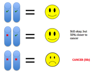
etic mutations and environmental exposures during one’s lifetime. Cancer is not heritable, but a predisposition to the disease can be (Peltomaki, 2012). For example, in our DNA we have two copies of each tumor suppressor gene, one from each parent. These genes keep cells from growing uncontrollably. So to lose function, one has to have mutations in both copies. Unfortunately, one can be born with a mutation in one copy, such as in Retinoblastoma (Rb). This means the gene is still expressed using the normal copy, but this confers a 50% increased predisposition for the disease bringing cells halfway closer to being cancer cells (Price et al., 2014).

Evolutionary Cancer Mutations

Misconception: It takes one bad mutation in one cell to get cancer, and cancer is one disease with all of cells in a tumor being equal.

Truth: Population biology is used to describe tumor growth and spread (metastasis), rather than arising from a single “bad” cell. Actually, it takes on average six to seven cumulative mutations (less in pediatric cancers) to confer disease, each one having been selected for during the previous cell generation. When mutations happen and build upon each other, the combination of changes can lead to cancer. (Yamamoto, Nakamura, & Haeno, 2015). Illustrated below. This supports cancer mostly being a disease of old age, as mutations take time to accumulate. Cancer essentially consists of many diseases that continue to harass the brains of researchers. Essentially, doctors have been able to treat for certain mutations, but once one important mutation is treated, another one can take over to drive relapse (Landau et al., 2015). Also, in tumors there ar2
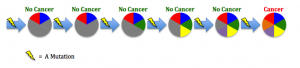
e different environments around cells fostering different mutations in various areas of the same tumor (Hardiman et al., 2015). Cancer is adapting to therapies that target mutations, making it so difficult to control.

DNA Damage

Misconception: DNA damage is rare, and we have little protection against it. Most cancer-causing agents (carcinogens) are processed chemicals not naturally found in nature.

Truth: DNA damage happens many times per day and our bodies have also evolved repair mechanisms in response to the need to correct such damage. In addition, the vast majority of cancer causin3
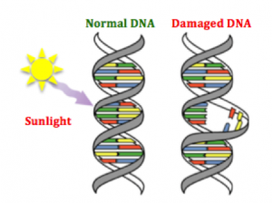
g agents are naturally occurring substances we encounter on a daily basis (Bauer, Corbett, & Doetsch, 2015). The human body has evolved ways to counteract DNA-damaging events, for example during sunlight exposure, using molecular machinery and likewise other naturally occurring compounds (Nishisgori, 2015).


References

Bauer, Nicholas C., Anita H. Corbett, and Paul W. Doetsch. “The Current State of Eukaryotic DNA Base Damage and Repair.” Nucleic Acids Res Nucleic Acids Research (2015): n. pag. Web.

Hardiman, Karin M., Peter J. Ulintz, Rork D. Kuick, Daniel H. Hovelson, Christopher M. Gates, Ashwini Bhasi, Ana Rodrigues Grant, Jianhua Liu, Andi K. Cani, Joel K. Greenson, Scott A. Tomlins, and Eric R. Fearon. “Intra-tumor Genetic Heterogeneity in Rectal Cancer.” Lab Invest Laboratory Investigation (2015): n. pag. Web.

Landau, Dan A., Eugen Tausch, Amaro N. Taylor-Weiner, Chip Stewart, Johannes G. Reiter, Jasmin Bahlo, Sandra Kluth, Ivana Bozic, Mike Lawrence, Sebastian Böttcher, Scott L. Carter, Kristian Cibulskis, Daniel Mertens, Carrie L. Sougnez, Mara Rosenberg, Julian M. Hess, Jennifer Edelmann, Sabrina Kless, Michael Kneba, Matthias Ritgen, Anna Fink, Kirsten Fischer, Stacey Gabriel, Eric S. Lander, Martin A. Nowak, Hartmut Döhner, Michael Hallek, Donna Neuberg, Gad Getz, Stephan Stilgenbauer, and Catherine J. Wu. “Mutations Driving CLL and Their Evolution in Progression and Relapse.” Nature 526.7574 (2015): 525-30. Web.

Nishisgori, Chikako. “Current Concept of Photocarcinogenesis.” Photochem. Photobiol. Sci. 14.9 (2015): 1713-721. Web.

Peltomäki, Päivi. “Mutations and Epimutations in the Origin of Cancer.” Experimental Cell Research 318.4 (2012): 299-310. Web.

Price, E. A., K. Price, K. Kolkiewicz, S. Hack, M. A. Reddy, J. L. Hungerford, J. E. Kingston, and Z. Onadim. “Spectrum of RB1 Mutations Identified in 403 Retinoblastoma Patients.” Journal of Medical Genetics 51.3 (2013): 208-14. Web.

Weinberg, Robert A. The Biology of Cancer. New York: Garland Science, 2007. Print.

Yamamoto, Kimiyo N., Akira Nakamura, and Hiroshi Haeno. “The Evolution of Tumor Metastasis during Clonal Expansion with Alterations in Metastasis Driver Genes.” Sci. Rep. Scientific Reports5 (2015): 15886. Web.


Evolution of Methicillin-Resistant Staphylococcus Aureus

Contributed by Priya Chopra, Shoeb Lallani, Rahul Mohan, Vivek Sawhney, Matt Wu, Manal Zafar

Perhaps you may think that humans cannot influence the evolution of MRSA. However, this is not the case. For years, humans have used antibiotics to treat many types of bacterial infections, ranging from Staphylococcus aureus infection to Streptococcus. Antibiotics can be found in places you might not expect, such as hand soaps, cleaners, toothpaste, and in livestock that have contact with humans (Nerby et al., 2011). The use of antibiotics in a wide variety of places promotes the development of a diverse range antibiotic resistance in S.aureus.

A Video on The Evolution of MRSA

You may also think that MRSA evolved/continues to evolve randomly by chance. However, the fact that humans continually introduce numerous antibiotics to bacteria promotes the evolution of bacteria in a way that promotes its own survival. Humans provide the selective pressure to prompt bacteria to evolve characteristics that will maximize its fitness in its host. For example, if we introduce an antibiotic to a population, there may be a select few bacteria that have a mutation or gene that confers antibiotic resistance, which can then be passed onto future generations (vertical gene transfer). This leads to antibiotic resistance, and, to make things worse, resistance can then also be transferred by horizontal gene transfer (between different species) (Giedraitiene et al., 2011). For example, S. aureus can develop resistance to an antibiotic used to treat a nearby different bacterial infection, like tuberculosis, by means of horizontal gene transfer. This resistance can then be shared to the rest of the S. aureus population via vertical gene transfer.

Eventually, many strains of MRSA evolve, and new antibiotics need to be created in order to treat the strains. So, next time your doctor gives you antibiotics, think about the evolutionary consequences of introducing these antibiotics to your body.

To learn more…

Cogen A. L., Nizet V., Gallo R. L. (2009). Skin microbiota: a source of disease or defense? British Journal of Dermatology, 158(3): 442-455.

Fomda B. A., Thokar M. A., Ray P. (2014). Prevalence and genotypic relatedness of methicillin resistant Staphylococcus aureus in a tertiary care hospital. Journal of Postgraduate Medicine, 60(4): 386-9.

Giedraitiene A., Vitkauskiene A., Naginiene R., Pavilonis A. (2011). Antibiotic Resistance Mechanisms of Clinically Important Bacteria. Medicina, 47(3): 137-46.

McNulty C., Boyle P., Davey P. (2007). The public’s attitudes to and compliance with antibiotics. Journal of Antimicrobial Chemotherapy, 60: 63-68.

Micek S. T. (2007). Alternatives to Vancomycin for the Treatment of Methicillin-Resistant Staphylococcus aureus Infections. Clinical Infectious Diseases, 45: 184-190.

Nerby J.M., Gorwitz R., Harriman K. (2011). Risk factors for household transmission of community-associated methicillin-resistant Staphylococcus aureus. Pediatric Infectious Disease Journal, 30(11): 927-32.

Planet P. J., LaRussa S. J., Dana A., Smith H., Xu A. (2013). Emergence of the Epidemic Methicillin-Resistant Staphylococcus aureus Strain USA300 Coincides with Horizontal Transfer of the Arginine Catabolic Mobile Element and speG-mediated Adaptations for Survival on Skin. American Society for Microbiology, 4, 13.

Poole K. (2007) Efflux pumps as antimicrobial resistance mechanisms. Annals of Internal Medicine, 39(3): 162-76.

Wielders C. L., Fluit A., Schmitz F., mecA Gene Is Widely Disseminated in Staphylococcus aureus Population, Journal of Clinical Microbiology, 40(11): 3970-3975.