Systematic Penguin Evolution

Contributed by: Oceana Hopkins, Arooj Khalid, Kevin Lu

The core idea of modern evolutionary theory is that all life is descended from a common ancestor. Though the theory garners much scrutiny and skepticism, it can be explained in part through the simple mechanism of natural selection. Natural selection takes advantage of the variability that exists within the genome. Random mutations that occur in the genome are behind these variations and sometimes change the fitness of an organism. Natural selection dictates that those with higher fitness survive and reproduce; thus, certain traits are selected for within the species. This process explains how gradual change occurs and how complex organisms arise from simplistic ancestors. A common misconception regarding evolution is that life evolved randomly, or by chance. This misconception could arise because of the random nature of mutations that promote variability. Though randomness is an important component to evolutionary theory, natural selection and survival of those who are more biologically fit make sure that the process is non-random.

Known for their characteristic black and white “tuxedos”, penguins are an aquatic, flightless bird found in both warm and cold climates. Because such a large part of their lives are spent in the water, certain species of penguins will only exit the water to shed their feathers or to mate. Spending nearly 75% of their lives in the water foraging for food, penguins have developed very specific traits optimized for swimming.  Utilizing their strong forelimbs to propel their large bodies through the water, penguins have undergone very specific evolutionary changes that allow this mechanism to run smoothly. As discussed in Michael Habib’s paper on structural evolution, the strength of the bones in the forelimbs are significantly greater than that of birds who do not exhibit aqua-flying behavior (2009). In conjunction with increased bone density, more muscle mass also developed and aided in keeping the penguins warm in their cold feeding environment.

Closer inspection of the features that aid water-feeding behavior provides more evidence that disputes the misconception that evolution is random. As seen in 2006 by Slack and colleagues, macroevolution within penguins based on their fossils and mitochondrial genes was tracked and recorded. The evidence showed that the penguins’ bodies gradually adapted to the cold feeding environment over many generations. Additionally, researchers Thomas and colleagues observed in 2010 that cold water penguins have a flow of heat along their wings that originates from the brachial artery called the humeral plexus.  This vascular countercurrent heat exchanger (CCHE) provides penguins the opportunity to forage in cold water by limiting heat loss through the flippers. Scientists identified this adaptation through fossil evidence and, upon further research, learned it evolved after penguins lost the ability of aerial flight. Researchers have proposed the CCHE evolved to help balance the energy costs of longer foraging times, since the oceans were significantly cooler than penguin body temperature. Ultimately, researchers have concluded that the humeral plexus was instrumental in allowing penguins to be water feeders in subaquatic environments.


To learn more:

Clarke, J. A., D. T. Ksepka, R. Salas-Gismondi, A. J. Altamirano, M. D. Shawkey, L. D’alba, J. Vinther, T. J. Devries, and P. Baby. “Fossil Evidence for Evolution of the Shape and Color of Penguin Feathers.”Science 330 (2010): 954-57. Print

Fordyce, R. E. and Jones, C. M. 1990. The history of penguins, and new fossil penguin material from New Zealand. Pages 419-446 in Davis, L. S. and Darby, J. D. (editors), Penguin biology. Academic Press, San Diego. 467 p.

Habib, Michael. “The Structural Mechanics And Evolution Of Aquaflying Birds.” Biological Journal of the Linnean Society 99 (2009): 687-98. Print.

Stack, Kerryn E., Craig M. Jones, Tatsuro Ando, G. L. Harrison, R. Ewan Fordyce, Ulfur Arnason, and David Penny. “Molecular Biology and Evolution.” Early Penguin Fossils, Plus Mitochondrial Genomes, Calibrate Avian Evolution. Oxford Journals, Mar. 2006. Web. 13 Nov. 2015.

Subramanian S, Beans-Pico´n G, Swaminathan SK, Millar CD, Lambert DM. 2013 Evidence for a recent origin of penguins. Biol Lett 9: 20130748. http://dx.doi.org/10.1098/rsbl.2013.0748.

Thomas, D. B., D. T. Ksepka, and R. E. Fordyce. “Penguin Heat-retention Structures Evolved in a Greenhouse Earth.” Biology Letters 7 (2010): 461-64. Print.

Evolution of Methicillin-Resistant Staphylococcus Aureus

Contributed by Priya Chopra, Shoeb Lallani, Rahul Mohan, Vivek Sawhney, Matt Wu, Manal Zafar

Perhaps you may think that humans cannot influence the evolution of MRSA. However, this is not the case. For years, humans have used antibiotics to treat many types of bacterial infections, ranging from Staphylococcus aureus infection to Streptococcus. Antibiotics can be found in places you might not expect, such as hand soaps, cleaners, toothpaste, and in livestock that have contact with humans (Nerby et al., 2011). The use of antibiotics in a wide variety of places promotes the development of a diverse range antibiotic resistance in S.aureus.

A Video on The Evolution of MRSA

You may also think that MRSA evolved/continues to evolve randomly by chance. However, the fact that humans continually introduce numerous antibiotics to bacteria promotes the evolution of bacteria in a way that promotes its own survival. Humans provide the selective pressure to prompt bacteria to evolve characteristics that will maximize its fitness in its host. For example, if we introduce an antibiotic to a population, there may be a select few bacteria that have a mutation or gene that confers antibiotic resistance, which can then be passed onto future generations (vertical gene transfer). This leads to antibiotic resistance, and, to make things worse, resistance can then also be transferred by horizontal gene transfer (between different species) (Giedraitiene et al., 2011). For example, S. aureus can develop resistance to an antibiotic used to treat a nearby different bacterial infection, like tuberculosis, by means of horizontal gene transfer. This resistance can then be shared to the rest of the S. aureus population via vertical gene transfer.

Eventually, many strains of MRSA evolve, and new antibiotics need to be created in order to treat the strains. So, next time your doctor gives you antibiotics, think about the evolutionary consequences of introducing these antibiotics to your body.

To learn more…

Cogen A. L., Nizet V., Gallo R. L. (2009). Skin microbiota: a source of disease or defense? British Journal of Dermatology, 158(3): 442-455.

Fomda B. A., Thokar M. A., Ray P. (2014). Prevalence and genotypic relatedness of methicillin resistant Staphylococcus aureus in a tertiary care hospital. Journal of Postgraduate Medicine, 60(4): 386-9.

Giedraitiene A., Vitkauskiene A., Naginiene R., Pavilonis A. (2011). Antibiotic Resistance Mechanisms of Clinically Important Bacteria. Medicina, 47(3): 137-46.

McNulty C., Boyle P., Davey P. (2007). The public’s attitudes to and compliance with antibiotics. Journal of Antimicrobial Chemotherapy, 60: 63-68.

Micek S. T. (2007). Alternatives to Vancomycin for the Treatment of Methicillin-Resistant Staphylococcus aureus Infections. Clinical Infectious Diseases, 45: 184-190.

Nerby J.M., Gorwitz R., Harriman K. (2011). Risk factors for household transmission of community-associated methicillin-resistant Staphylococcus aureus. Pediatric Infectious Disease Journal, 30(11): 927-32.

Planet P. J., LaRussa S. J., Dana A., Smith H., Xu A. (2013). Emergence of the Epidemic Methicillin-Resistant Staphylococcus aureus Strain USA300 Coincides with Horizontal Transfer of the Arginine Catabolic Mobile Element and speG-mediated Adaptations for Survival on Skin. American Society for Microbiology, 4, 13.

Poole K. (2007) Efflux pumps as antimicrobial resistance mechanisms. Annals of Internal Medicine, 39(3): 162-76.

Wielders C. L., Fluit A., Schmitz F., mecA Gene Is Widely Disseminated in Staphylococcus aureus Population, Journal of Clinical Microbiology, 40(11): 3970-3975.  

Helicobacter pylori and you.

Contributed by Thomas Partin and Austin Piccolo

Species do not live in a world separate from each other. Organisms interact with other organisms everyday, and over time adapt to each other accordingly. Often, the evolution of two species can become strongly linked to each other, for better or worse. Heliobacter pylori is a bacteria that thrives in the acidic conditions of the human stomach. It causes stomach ulcers and is strongly correlated with gastric cancer. As recently as the 1980’s, the idea of a bacteria being able to survive in the stomach’s harsh conditions and being responsible for this disease was so controversial that it took one doctor intentionally infecting himself to prove its role in stomach ulcers. That doctor later won the Nobel prize in medicine for his work.

H. pylori did not first start infecting humans in the 80’s though. H. pylori and humans have been living (and battling) together for millennia. The earliest humans also played host to H. pylori. One way this can be shown is a creative use of a phylogenetic tree. Scientists sampled many different strains of H. pylori, and used them to create an ancestral tree of the different strains. They then compared the tree they made to geographic locations of their samples. What they found was that lineages of H. pylori matched perfectly with the migration patterns of ancient humans as they moved out of Africa. Newer strains of H. pylori are found where humans migrated to most recently. The strains were carried and dispersed based on how early humans moved around the globe.

This intimate relation between H. pylori and humans provides a great opportunity to explore coevolution. Humans and H. pylori have been locked in an arms race for thousands of years. H. pylori colonization poses serious health consequences to the host, which creates a selective pressure for humans that can prevent H. pylori infection. Likewise, the human body is an incredibly hostile environment towards foreign invaders like H. pylori, which creates a strong selective environment for H. pylori cells that can overcome human defenses. There is evidence this selective pressure is so strong that H. pylori begins adapting specifically to the host after initial colonization. Although not an innate aspect of human biology, antibiotics are another human defense against H. pylori. Antibiotic use creates a selective pressure for H. pylori that is so strong that resistant strains can develop remarkably quickly after attempted treatment.

Please watch the below video to learn more!

https://www.youtube.com/watch?v=01NY55VV0Rg;feature=youtu.be

For further information see:

Linz, B., Ballouxm, F., Moodley, Y., Manica, A., Liu, H., Roumagnac, P., Falush, D., Stamer, C., Prugnolle, F., van der Mer, S.W., Yamaoka, Y., Graham, D.Y., Perez-Trallero, E., Wadstrom, T., Suerbaum, S., Achtman, M. 2007. An African origin for the intimate association between humans and Helicobacter pylori. Nature 445: 915-918

Gao, W., Cheng, H., Hu, F., Li, J., Wang, L., Yang, G., Xu, L., Zheng, X. 2010. The Evolution of Helicobacter pylori Antibiotics Resistance Over 10 Years in Beijing, China. Helicobacter. 15: 460-466.

Oh, J.D., Kling-Bäckhed, H., Giannakis, M., Xu, J., Fulton, R.S., Fulton, L.A., Cordum, H.S., Wang, C., Elliott, Glendoria., Edwards, J., Mardis, E.R., Engstrand, L.G., Gordon, J.I. 2006. The complete genome sequence of a chronic atrophic gastritis Helicobacter pylori strain: Evolution during disease progression. PNAS. 103: 9999-10004.

Blecker, U., Landers, S., Keppens, E., Vandenplas, Y. 1994. Evolution of Helicobacter pylori Positivity in Infants Born From Positive Mothers. Journal of Pediatric Gastroenterology and Nutrition. 19: 87-90

Kennemann, L., Didelot, X., Aebischer, T., Khun, S., Drescher, B., Droge, M., Reinhardt, R., Correa, P., Meyer, T.F., Josenhan, C., Falush, D., Suerbaum S. 2011. Helicobacter pylori genome evolution during human infection. PNAS. 108: 5033-5038.

Marshall, B.J., Warren, J.R. 1984. Unidentified curved bacilli in the stomach of patients with gastritis and peptic ulcers. The Lancet. 323: 1311-131.

Avasthi, T.S., Devi, S.H., Taylor, T.D., Kumar, N., Baddam, R., Kondo, S., Suzuki, Y., Lamouliatte, H., Mégraud, F., Ahmed, N. 2011. Genomes of Two Chronological Isolates (Helicobacter pylori 2017 and 2018) of the West African Helicobacter pylori Strain 908 Obtained from a Single Patient. Journal of Bacteriology. 193: 3385-3386.

The Lilliput Effect: Trends in Body Size Following Ancient Mass Extinctions

Contributed by Ziqi Wu, Tian Mi, Lei Huang, Zhuo Li.

Imagine myriad fishes, including creatures the size of school buses, swimming in the Earth’s seas. Yup. That’s how things used to be 360 million years ago. It was not until the appearance of Devonian-Mississippian vertebrates that the modern range of body sizes became readily visible in the marine system. What happened in between these times? What made the creatures’ sizes shrink and spoiled the fun of scuba diving nowadays?

The answer is that a massive extinction happened about 359 million years ago, at the end of the Devonian Period. The scientists named it the Lilliput Effect: a temporary size reduction following mass extinction, which is usually temporary, but sometimes becomes persistent in specialized groups, such as birds, plankton, or island faunas. But how can a smaller size increase an organism’s fitness? One popular misconception is that the fittest organisms in a population are those that are strongest and largest. However, the Lilliput Effect has proved this wrong.

cartoon

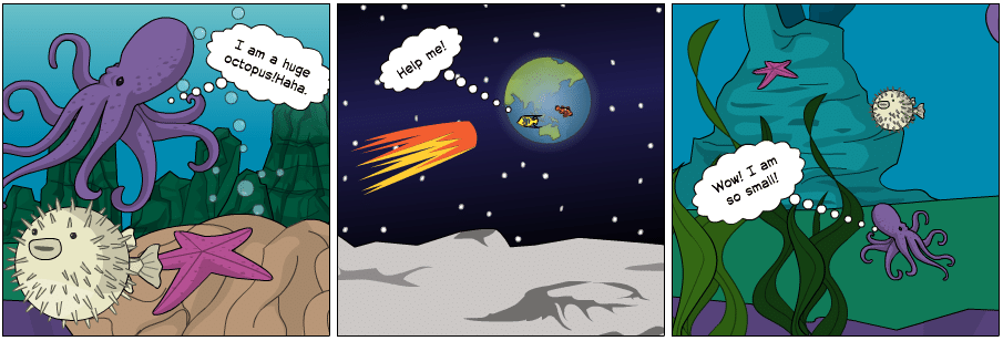
A giant fish has the length of a school bus before the die-off. However, after the asteroid hit earth and a mass extinction took place, the surviving fishes, and their descendants, are smaller since smaller size gives them an advantage.

The mechanism underlying the effect is controversial. One hypothesized model points to the effect of weather. Researchers (Dahl et al.) tracked down redox history of the atmosphere and oceans and found out that the radiation of large predatory fish (animals with high oxygen demand) correlates with atmosphere oxygenation. Therefore, a lower level of oxygen in the atmosphere might have played a role in the overall shrinkage. A temperature model based on Bergmann’s rule proposes that size is negatively influenced by temperature (Bergmann 1847). Previous studies suggested that warm-blooded vertebrate species are larger in colder climates than their congeners inhabiting warmer climates (Harries and Knorr 2009).

However, a more recent study points to another advantage of smaller size: smaller individuals grow and reproduce faster, and they adapt to their environments more quickly because of their relatively short generation times (Sallan and Galimberti 2015). Using holocephalans and ray-finned fishes as examples, they demonstrated that smaller vertebrates tend to have high reproductive rates, short generation times, and large populations. These traits may increase survival while promoting diversification via higher variation and population fragmentation.

Altogether, although there is empirical evidence suggesting that Lilliput effect could be a general response to environmental stress following various mass extinction events, there remain many puzzles to be solved.

For further reading, see:

Adam K. Huttenlocker and Jennifer Botha-Brink. 2013. Body size and growth patterns in the therocephalian Moschorhinus kitchingi (Therapsida: Eutheriodontia) before and after the end-Permian extinction in South Africa. Paleobiology 39(2), 353-277.

Bing Huang, David A.T. Harper, Renbin Zhan, Jiayu Rong. 2010. Can the Lilliput Effect be detected in the brachiopod faunas of South China following the terminal Ordovician mass extinction? Palaeogeography, Palaeoclimatology, Palaeoecology, Volume 285, Issues 3–4, Pages 277-286.

Keller, G., Abramovich,S. 2009. Lilliput effect in late Maastrichtian planktic foraminifera: Response to environmental stress. Palaeogeography, Palaeoclimatology, Palaeoecology 284: 47-62

Peter J. Harries, Paul O. Knorr. 2009. What does the ‘Lilliput Effect’ mean? Palaeogeography, Palaeoclimatology, Palaeoecology, Volume 284, Issues 1–2, Pages 4-10.

Richard J. Twitchett. 2007. The Lilliput effect in the aftermath of the end-Permian extinction event, Palaeogeography, Palaeoclimatology, Palaeoecology, Volume 252, Issues 1–2, Pages 132-144, ISSN 0031-0182.

Sallan L, Galimberti AK. 2015. Body-size reduction in vertebrates following the end-Devonian mass extinction.Science 350(6262):812-815.


Climate Change and Evolution

Contributed by Sarah Meadows

Climate change has the potential to shape evolutionary change in species, and has already done so in a few species around the planet.  Climate change, or global warming as it is also called, can exact selectional pressure on species because of how it changes the habitat. Changing temperatures is perhaps the most well-known effect of climate change. Climate change can exert selectional pressure because not all species are fit enough to survive when their habitat changes.

One of the challenges with studying climate change and evolution is that it can be hard to distinguish between plastic responses and genetic changes. Plasticity is a species’ ability to adapt to changing conditions without their genetics, or their DNA, being changed. If a species breeding time is earlier than before and there is a correlation with warmer temperatures, for example, it could be because most individuals have the ability to begin breeding earlier (a plastic response), or it could be because individuals that happen to breed earlier have higher survival and reproduction, and they pass this early breeding trait onto their offspring. The latter scenario represents evolution by natural selection. Much of the research done so far on the subject has measured a change in life history traits but has not determined if the cause is a genetic change or phenotypic  plasticity.

Species can respond to climate change in a few ways. They can change their life history traits, they can expand or shift their range, or they can go extinct. The latter has already happened to some amphibian populations, such as the golden toad and Monteverde harlequin frog in Costa Rica.

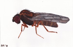
Some species that have been studied extensively on the subject are Drosophila flies, Darwin’s finches, and many other bird species. Some Drosophila have been found to exhibit genetic changes in the northern ranges that are very similar to ones from their southern ranges, suggesting that they either migrated northwards or there was directional selection for those genes.  Some other changes that have been observed in other species due to climate change are ranges moving northwards, egg laying and breeding periods happening earlier, and migration happening later.

fly

A few species of Drosophila have recently experienced genetic changes that have been attributed to climate change.

Even though some species have the ability to respond to climate change, such as adapting to the new conditions or moving, not all species can do so.  Amphibians, for example, are hit very hard by it, with high rates of extinction compared to other vertebrates.  Coral reefs are also hit hard by climate change because higher CO2 levels and bleaching (losing their microbial partners) is a problem.  Another problem that is posed by climate change is that if two species have coevolved and are symbiotic with each other, one species could experience evolution due to climate change and the other may not.  For example, a pollinating insect could emerge too early before the plant species it usually pollinates has bloomed.

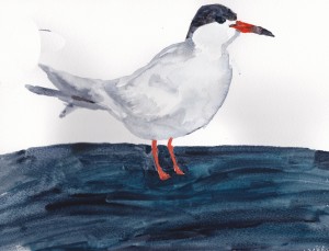
birdy

Artic terns are one species of birds whose breeding date has advanced, possibly because of climate change. This is an example of a phenotypic change.

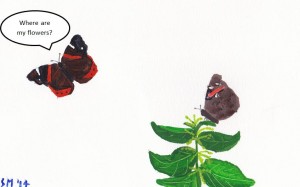
bugs

Insects can be affected by asynchrony, for example when a butterfly hatches earlier then its host plant. 

Climate change presents a new, challenging influence on habitats, and its long term effects on species diversity and their evolution have yet to be determined, but in the short term it is clear that climate change does have an effect on the evolution of species.

For more information, see:

Balanyá, J., Oller, J. (2006) Global Genetic Change Tracks Global Climate Warming in Drosophila subobscura. Science. 313: 1773-1775.

Gienapp, P., Teplitsky, C., Alho, J. S., Mills, J. A. and Merila, J. (2008), Climate change and evolution: disentangling environmental and genetic responses. Molecular Ecology, 17: 167–178.

Grant, P., Grant, R. (1995), Predicting microevolutionary responses to directional selection on heritable variation . Evolution, 49: 241-251.

Levitan, M., Etges, W., (2005) Climate change and recent genetic flux in populations of Drosophila robusta. BMC Evolutionary Biology, 5:4

MØller, A. P., Flensted-Jensen, E. and Mardal, W. (2006), Rapidly advancing laying date in a seabird and the changing advantage of early reproduction. Journal of Animal Ecology, 75: 657–665.

Parmesan, C. (2006), Ecological and evolutionary responses to recent climate change. Annual Review of Ecology Evolution and Systematics,  37: 637-669.

Pounds, A., Bustamante, M. (2006), Widespread amphibian extinctions from epidemic disease driven by global warming.  Nature, 439: 161-167

The Evolution of Bitter Taste

Contributed by Jonathan Adcock, Hana Ahmed, Robert Bruner, Farhan Momin, Andrew Shibata

How Bitter Taste Works

Specialized bitter taste receptors are concentrated at the back of the tongue. Upon eating a bitter food, these receptors are activated, and and a signal is sent to to the brain that leads to the perception of a bitter taste. Bitter taste receptors are encoded by the TAS2R gene family. This family includes nearly 25 genes and psuedogenes (genes that are no longer functioning) that are concentrated in bundles on chromosomes 3, 5, and 7. Extensive studies have been performed in order to determine which molecules in bitter foods can activate these receptors. Scientists have found that many of the compounds that activate the bitter taste receptors are chemicals produced by plants. Many of these compounds were also found to be toxic and if consumed could lead to illness or death.

How Bitter Taste Evolved

Our ancestors and other animals have not always been able to taste bitter foods. The bitterness sensation is thought to have evolved 200 million years ago. The prevalent hypothesis is that bitter taste evolved by random gene mutation events which caused the formation of the TAS2R gene family and the bitter taste receptors on the tongue that could bind to toxic chemicals. Animals possessing these mutations were able to  taste toxins in their food. These toxins would have tasted bad to the animal, and thus, the animal would learn to avoid the toxic food in the future. Animals with the mutations that produce the TAS2R gene family would be be better adapted to their environment because they can avoid toxic and poisonous foods that could cause sickness or death in the animal. Animals not possessing these mutations would still be susceptible to ingesting naturally occurring poisonous chemicals. Because these animals have improved survivability in comparison to other animals without the TAS2R genes, animals that could taste bitter foods would be better able to reproduce and pass the functional TAS2R genes to their offspring, who in turn would have increased survivability, be better able to reproduce and pas the TAS2R genes to their offspring. Organisms possessing the TAS2R genes have a higher fitness than organisms that do not posses this gene family. Thus, the TAS2R gene family would have been selected for via natural selection and became the ability to taste bitter foods would have become dominant in the population.

This is especially true in the case of the thyroid-inhibiting toxin, PTC. PTC is known to decrease thyroid hormone, which regulates metabolic function. PTC can also lead to liver damage and a host of other medical problems that would eventually lead to death. Being able to taste the PTC toxin would have allowed our ancestors to to avoid toxic foods that if ingested would ultimately lead to their death. Because of this, the ability to taste the PTC toxin would be passed on to the next generation and contribute to the increased fitness of the following generation. In this way, the bitter taste genes would become selected for and would become prevalent in the population.

How the Evolution of Bitter Taste Affects Me Today

Although variation in taste facilitated the evolution of primates and other animals, in modern human society it can be detrimental. With the advent of agriculture, there is less need for tasting of bitter food. Highly nutritious vegetables are known to activate the bitter taste receptors. Instead of doing their intended evolutionary job of protecting from toxic materials, the bitter taste receptors are preventing some people from getting an adequate daily amount of nutritious vegetables due to their aversive taste.

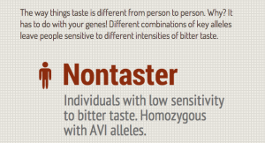
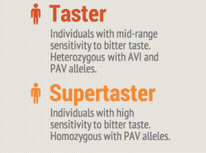
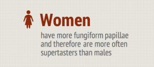
Additionally, some individuals have extremely high sensitivity to bitter tastes resulting from higher rates of expression of the TAS2R gene. These individuals are known as “supertasters”.  Supertasters can be found in the highest frequency in parts of Asia, Africa, and South America. This may be because these areas originally possessed a higher concentration of toxic plants and animals in comparison to other areas. In order to compensate for the increase in potential toxins, the individuals living in this area might have benefited more from bitter taste receptors. Today, many supertasters dislike a wide variety of vegetables such as cabbage and soy, and favor sweet items over bitter vegetables. This not only leads to increased chance of obesity and diabetes, but because of the avoidance of vegetables there is increased risk for GI diseases such as colon cancer. Not all side effects are bad though. Supertasters have a higher dislike for alcohol, carbonated beverages, and smoking. Avoidance of these items is actually quite beneficial in boosting heath.

Next time you don’t feel like eating your vegetables, you can blame it on evolution!

 Screen Shot 2014-04-29 at 3.28.53 PM

Screen Shot 2014-04-29 at 3.29.16 PM Screen Shot 2014-04-29 at 3.29.37 PM Screen Shot 2014-04-29 at 3.30.09 PM Screen Shot 2014-04-29 at 3.30.23 PM
More Reading…

Campbell, M. C., P. A. S. Breslin, A. Ranciaro, S. A. Tishkoff, D. Drayna, D. Zinshteyn, G. Lema, T. Nyambo, J.-M. Bodo, S. Omar, J. Hirbo, and A. Froment. “Evolution of Functionally Diverse Alleles Associated with PTC Bitter Taste Sensitivity in Africa.” Molecular Biology and Evolution 29.4 (2012): 1141-1153.

Wooding, Stephen. “Evolution: A Study in Bad Taste?.” Current Biology 15.19 (2005): R805-R807.

Human Impact on Altered Behavior and Evolution of Species

Contributed by Brad Richardson

A common misconception about evolution is that it is only a theory; however, it is a fact. A fact is a truth known by actual experience or observation. Darwin’s Theory of Evolution by Natural Selection on the other hand is a theory much like the theory of relativity or gravity; it is a broad explanation of a particular phenomena that is proposed based on multiple observations and can be used to make predictions.

Many believe the theory is not testable or observable and that if it is true, it only happens over a very long period of time. However, human intervention via our technological advances such as city building and industrialization has led to an altered environment that is changing at an extreme rate. Although this can have either negative or positive consequences for varying species, one positive consequence for the human species is that it is allowing us to get a much better understanding of the scope and specificity of how evolutionary mechanism works.

Because of this accelerated rate of technological advance by humans, others species are having to adapt and evolve at an equally fast rate. This allows us to get a unique glimpse of evolution happening before our eyes. One of the biggest culprits is of course the effect climate change is having on all species, including our own. Various animals are breeding earlier in the spring, becoming smaller to keep their body temperatures in balance, and even completely moving their ranges away from the equator to avoid the heat!

Many changes are also occurring at the microscopic level due to technological advances in medicine. Antibiotic resistant bacteria have evolved to increase their chances of survival. The bacteria evolve and reproduce at such a fast scale that “superbugs” even exist which evolve resistance to the antibiotics that are usually given as an alternative to regular antibiotics.

Changes are happening all around us, which makes the Theory of Evolution such an interesting topic to study because every day species are changing and adapting and providing “natural” observable data that further bolsters the legitimacy of the theory.

Take a look below at some examples of observable evolution!

https://www.youtube.com/watch?v=fioFFtvEXBw

For more information check out:

Candolin U, Nieminen A, Nyman J. 2014. Indirect effects of human-induced environmental change on offspring production mediated by behavioral responses. Oecologia. 174: 87-97.

Janssens L, Khuong DV, Debecker S, Bervoets L, Stoks R. 2014. Local adaptation and the potential effects of a contaminant on predator avoidance and antipredator responses under global warming: a space-for-time substitution approach. Evolutionary Applications. 7: 421-430.

Sol D, Lapiedra O, Ganzalez-Lagos C. 2013. Behavioural adjustments for a life in the city. Animal Behaviour. 85: 1101-1112.

Nemeth Z, Bonier F, MacDougall-Shackleton SA. 2013. Coping with uncertainty: integrating physiology, behavior, and evolutionary ecology in a changing world. Integrative and Comparative Biology. 53: 960-964.

Martin J, Lopez P. 2013. Effects of global warming on sensory ecology of rock lizards increased temperatures alter the efficacy of sexual chemical signals. Functional Ecology. 27: 1332-1340.

Miranda AC, Schielzeth H, Sonntag T, Partecke J. 2013. Urbanization and its effects   on personality traits: a result of microevolution or phenotypic plasticity?. Global Change Biology. 19: 2634-2644.

Evolution in Influenza

Contributed by Runako Aranha-Minnis

Evolution is not only limited to the organisms we are able to see with the naked eye. Viruses like influenza are able to evolve, and this can be very dangerous to human populations. Though it is true that influenza is not the danger that it once was, it still can not be ignored. Viral resistance to treatment can lead to many deaths globally, especially during flu seasons. During these times, doctors usually see a rise in cases of viruses resistant to the treatments available at the time. One type of resistance that influenza can develop is Oseltamivir resistance. Oseltamivir is an important antiviral defense against the flu. It is found in drugs like Tamiflu, and can be rendered ineffective if resistance is developed.

Influenza and its neuraminidase

The DNA change that in the virus that occurs is related to the neuraminidase enzyme activity. The enzyme cleaves salicylic acid moieties that can be bound by the viral hemagglutinin. In essence the enzyme’s job is to help release newly formed viral particles. Imagine the enzyme as little Pac-Men just going about and eating the connections of new virus particles to let them spread. Oseltamivir works by inhibiting the activity of the neuraminidase enzyme. This limits the infectiousness of the virus.

Resistance requires changes in the DNA of the virus. Mutations, changes in the DNA, can change genes, often leading to changes in the organism. For Oseltamivir resistance, a point mutation, the change of a single base of DNA, is all that is necessary. A change in the genes leads to a change in the amino acids produced. This change in the amino acids is just one amino acid at the 274th spot in the chain leads to a change in the activity of the virus. This line of reasoning is why the mutation is called H274Y.

The H274Y mutation that gives influenza resistance usually does not spread through the population.  In early clinical tests, the mutation meant the virus was unaffected by oseltamivir, but had associated decreased viral fitness. So even though viruses with resistance would be selected for, their lower chance of survivability because of the mutation meant it did not matter. The influenza virus is able to evolve with Oseltamivir only when an associated mutation occurs. These associated mutations that allow oseltamivir resistance to keep going are permissive mutations. More recently, permissive mutations have arisen that allow influenza to be resistant to Oseltamivir without losing any fitness. Imagine a population of influenza like a game of “Where is Waldo?”:

No permissive mutations. In the absence of permissive mutations, all viruses, including Waldo, die. 

With permissive mutations. If Waldo has a permissive mutation, he survives to reproduce and the whole populations becomes Waldos because all other viruses died. 

Waldo in the first cartoon has H274Y with no permissive mutations. Without permissive mutations, there is no evolution under treatment. After treatment, the viruses are killed. There are no viruses left so the picture goes black. In the second cartoon, With permissive mutations, there is evolution for Waldo and a new viral population arises. This population only has Waldo because only Waldo has the H274Y mutation that conferred resistance.

For Further Reading:

Jesse D. Bloom, Lizhi Ian Gong, David Baltimore. 2010. Permissive Secondary Mutations Enable the Evolution of Influenza Oseltamivir Resistance Science. 328: 1272-1275

Nicholas Renzette, Daniel R. Caffrey, Konstantin B. Zeldovich, Ping Liu, Glen R. Gallagher, Daniel Aiello, Alyssa J. Porter, Evelyn A. Kurt-Jones, Daniel N. Bolon, Yu-Ping Poh, Jeffrey D. Jensen, Celia A. Schiffer, Timothy F. Kowalik, Robert W. Finberg, Jennifer P. Wang. 2013. Evolution of the Influenza A Virus Genome during Development of Oseltamivir Resistance In Vitro 88: 272-281

Superbugs: The Evolution of Gonorrhea

Contributed by Nyasia Jones, Chris Richardson and Kari Tyler

https://www.youtube.com/watch?v=AesE046gtQs

A common misconception regarding evolution it that it is slow, and because it is slow, humans are not influencing it. This is completely false. Humans have and are continuing to make major changes that are not only influencing the course of our own evolution, but are also influencing the evolution of other species we interact with. Especially in medicine, human advancement is occurring at an amazing pace and thus allowing us to witness evolution in response to our actions.

Over the past century, the use of antibiotics to treat bacterial pathogens has become a widespread practice. Starting around 1930, medical practitioners began discovering several drugs that successfully rid patients of a particular bacterial pathogen Neisseria gonorrhoeae. The first successful therapy was administration of a class of chemicals called sulfonamides. For years, treatment with sulfonamides proved successful in patient after patient until about the mid-1940s when reports of N. gonorrhoeae resistance to sulfonamides increased. Fortunately, at this time, the “miracle drug” penicillin was found to be highly effective at bacterial treatment and become the number one treatment option. After ten to fifteen years though, low doses of penicillin were no longer as effective, and by the 1980s strains with high-level penicillin resistance had emerged.

So what happened with both penicillin and the sulfonamides?  Nothing.

But something did happen to the N. gonorrhoeae. They evolved.

Sequential chromosomal mutations allowed some bacteria to incur resistance to penicillin. With penicillin treatment, the bacteria with resistance survived while those without it did not. The resistant individuals reproduced thereby creating a new generation of bacteria in which all individuals were penicillin resistant. This, my friends, is evolution.

Unlike the billions and billions of years it took to create modern-day humans, this evolution took less than a century to change these N. gonorrhoeae bacteria into “superbugs” which are becoming increasingly harder to treat. And it doesn’t stop there. N. gonorrhoeae has also become resistant to more recent treatment options, such as tetracycline and fluoroquinolones. Now, with less and less success to current methods of treatment, namely cefixime and ceftriaxone, scientists are worried history will repeat itself and strains of N. gonnorhoeae with complete cefixime and ceftriaxone resistance will emerge. With dwindling options for treatment, N. gonorrhoeae resistance and that of other superbugs remains a major problem in the fields of medicine and epidemiology.

So are humans influencing evolution? N. gonorrhoeae tell us, loud and clear: yes!

For more information:

Anderson, M. T., & Seifert, H. S. (2011). Neisseria gonorrhoeae and humans perform an evolutionary LINE dance. Mobile Genetic Elements, 1(1), 85-87.

Mavroidi, A., Tzelepi, E., Siatravani, E., Godoy, D., Miriagou, V., & Spratt, B. G. (2011). Analysis of emergence of Quinolone-resistant gonococci in Greece by combined use of Neisseria gonorrhoeae multiantigen sequence typing and multilocus sequence typing. Journal of Clinical Microbiology, 49(4), 1196-1201.

Unemo, M., & Shafer, W. M. (2011). Antibiotic resistance in Neisseria gonorrhoeae: Origin, evolution, and lessons learned for the future. Annals of the New York Academy of Sciences, 1230(1), E19-E28.

Venom Variation

Contributed by Annelise Bonvillian, Cherisma Patel and Liz Pinkerton

I introduce to you two rattlesnakes. I think they’re generally friendly but they could also be planning to maim me. You just never know with venomous snakes. 

Judging by their appearance, you’d think both rattlesnakes were of the same species. You’d be right, being the clever person you are. They are in fact part of the same subspecies of Southern Pacific Rattlesnake (Crotalus oreganus helleri).

These C. o. helleri rattlesnakes are venomous. Before we dive into their story, let’s explain venom.

Venom Across The Tree of Life

Many animals have evolved venom for protection or to help them capture prey. We, unfortunately have not.

There are also a wide range of structures for delivering venom, such as the cute little fangs in these rattlesnakes.

The interesting thing is, similar types of venom have evolved in completely unrelated species. This is not because all venomous species evolved from one super venomous ancestor or because the venomous critters of the world get together to talk strategy.

Evolution of venom is a solid example of convergent evolution in which organisms that are not very closely related independently evolve similar traits due to similar environmental pressures. The proteins in the toxin end up acting on the same physiological molecule by chance. Most venoms involve disrupting major physiological pathways that are accessible by the bloodstream, especially the hemostatic and neurological systems. Damaging these systems would efficiently cripple any prey or potential predator.

Back to the Rattlesnakes

Getting back to our original C. o. helleri rattlesnakes, you’d think it might be true that they’d have the same type of venom because they’re of the same subspecies, right?

In fact, these two rattlesnakes are very closely related, but the populations that live in the mountains have a distinctly different venom than those that live in the desert.

Distribution of Crotalus oreganus

Distribution of Crotalus oreganus

A research team from the University of Queensland studied the variation between rattlesnakes in the mountainous area of Idyllwild, California and those in the desert of Phelan, California. The extent of venom variation is surprising for two populations because the two areas are relatively close together geographically.

The venom of the desert-dwelling rattlesnake contains proteins that break down blood vessels and prevent clotting.

This venom is slow acting. It is described as haemotoxic and would lead to uncontrollable bleeding. Neat!

Its close relative in the mountains is equally as morbid. The mountain populations have proteins in their venom that stop nerves from sending signals to the muscles.

This venom is fast acting. It is described as neurotoxic and results in paralysis. Sweet!

The venoms are completely different even though these two populations are from the same subspecies.

In fact, venom can vary between species, subspecies, individuals, sexes, and ages. Another research team from the Biomedical Institute of Valencia that loves poisonous reptiles just as much as the the previous one studied venom variability in Mojave rattlesnakes in Arizona. They found that you were 10 times more likely to die from a bite in one county than another. Here, the severity, not the type, of venom was what varied.

So, getting back to our C. o. helleri rattlesnakes (don’t worry, we love you best), here’s another question: which would be better to have in the snake’s arsenal? The fast acting neurotoxin venom or the slow acting haemotoxin?

Evolution, the lovely process that developed these fine creatures is NOT about having the best of a trait. The fitness of a certain trait is relative. What is good for one population might not necessarily be the best for another. Based on where they live, different venom types might be more beneficial in capturing prey or warding off predators.

So why would it be more beneficial for the mountain rattlesnake to have a faster acting venom?

Researchers suspect that environmental differences between these two populations of rattlesnake are likely to have promoted the huge variation in venom between the two.

If the mountain rattlesnake’s venom was slow acting the, prey could hide before the venom had properly incapacitated it.

And venom isn’t cheap. Creating venom costs a lot of energy and wasting it time and time again would be a shame. Natural selection would thus lead to a venom composition that would reduce metabolic cost. Natural selection is the process by which organisms that have higher fitness and are more adapted to their environment tend to produce more offspring and survive.

Now, in the desert, rattlesnakes don’t have to worry as much about their prey hiding before they can get to them. They might not have evolved the quicker neurotoxic venom because there was no selective pressure to have their venom be fast acting.

As with most things in this incomprehensible universe, we are not sure if the desert dwelling Southern Pacific rattlesnake maintains its haemotoxic venom due to lack of selection pressure. Or perhaps somehow the snakes have co-evolved with their prey and the haemotoxic venom is just the most efficient one for the prey type.

All in all, it’s important to realize that evolution isn’t a race where the end goal is to have the best of something. Traits change randomly due to mutations, and factors such as the environment or prey type select for variations that are most beneficial for survival.

In related news, using the vast powers of human intelligence to learn from the natural world, snake venom is being adapted to heal instead of hurt. Venom works in the same way as many medicines, and the enzymes in venom are being modified to affect disease processes.

One specific example includes the using ACE inhibitors in Brazilian pit viper venom to prevent hypertension.

However, with the decline of snake diversity due to environmental degradation, the diversity of venom and its medical potential is decreasing.The fact that venom from various snakes can be used to target certain  diseases is a very important implication for evolutionary medicine. Population divergence in snakes increases the potential for variation in venom type, which can ultimately increase the antidotes possible for fighting certain diseases.

So why research things like rattlesnake venom? Not only is the subject wildly fascinating, but unraveling the complexities of snake venom can help humans better counter its life-threatening effects and can also promote the development of new medicines. In conclusion,  though we may be terrified of you, dear rattlesnakes and other venomous denizens of this world, we’d also like to say thanks. May you continue to amaze us.

Check out these sites for more information:

Caswell, Nocholas R., Wolfgang Wuster, Freek J. Vink, Robert A. Harrison, and Bryan G. Fry. 2012. Complex cocktails: the evolutionary novelty of venoms Trends in Ecology and Evolution.

Holland, Jennifer S. 2013. Venom: The Bite That Heals. National Geographic: The New Age of Exploration. http://ngm.nationalgeographic.com/2013/02/125-venom/holland-text

Kartik Sunagara, Eivind A.B. Undheimc, Holger Scheibd, Eric C.K. Grene, Chip Cochrane, Carl E. Persone, Ivan Koludarovc, Wayne Kellne, William K. Hayese, Glenn F. Kingd, Agosthino Antunesa, Bryan Grieg Fry. Intraspecific venom variation in the medically significant Southern Pacific Rattlesnake (Crotalus oreganus helleri): Biodiscovery, clinical and evolutionary implications. 2014. Journal of Proteomics. http://www.sciencedirect.com/science/article/pii/S1874391914000256

Massey DJ, Calvete JJ, Sánchez EE, Sanz L, Richards K, Curtis R, Boesen K. 2012. Venom variability and envenoming severity outcomes of the Crotalus scutulatus scutulatus (Mojave rattlesnake) from Southern Arizona. Journal of Proteomics. http://www.ncbi.nlm.nih.gov/pubmed/22446891

Yong, Ed. 2014. Rattlesnakes Two Hours Apart Pack Totally Different Venoms. National Geographic: Phenomena. Online. http://phenomena.nationalgeographic.com/2014/01/27/rattlesnakes-two-hours-apart-pack-totally-different-venoms/

Zimmer, Carl. 2013. On the Origin of Venom. National Geographic: Phenomena. Online. http://phenomena.nationalgeographic.com/2013/01/09/on-the-origin-of-venom/