One Large Plague Against Humanity, One Small Step Towards Immunity

By: Linda Huang, Shivani Seth, Shawn Kripalani, Anasua Bandyopadhyay and Nikita Maddineni

Imagine you’re living in the middle ages welding steel swords, leading your mundane life. Suddenly, your child is overcome with a horrid fever. Distraught, you also start to notice your child developing shockingly large lumps all over his body. What is going on? Soon, you learn that this nightmare is not only endangering your son’s life, but also the communities around you. Two weeks later, everyone around you has passed away, but for some reason you managed to survive. Why?

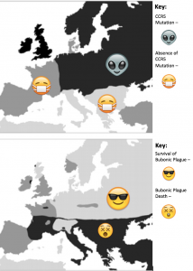
This so-called curse was actually known as the Black Plague that spread throughout Europe. The black death killed 75 to 200 million people and it peaked in Europe between the years of 1346 and 1353. It began with a rat infected with the bacterium Yersinia pestis. The bacteria infect humans by attacking the lymph nodes. They then begin to rapidly replicate, causing the lymph nodes to swell and become buboes. These buboes cause the immune system to go into septic shock, which is multiple-organ failure. This was one of the darkest points of European history, as 30-60% of Europe’s total population was killed. The methods of treatment ranged from visiting witch doctors to bathing in urine to turning to the church. Communities even started to live in the sewage systems after they became aware that it could be airborne. However, there was a small percentage of the population that was able to survive the plague.  In the population that survived, researchers found that there was a mutation in the gene for the CCR5 cell membrane receptor called CCR5-∆32.

This is an example of natural selection, where the bubonic plague acted as the selection pressure for individuals with the mutation. These individuals with the delta-32 mutation had a higher fitness because they were able to survive and reproduce better compared to individuals who were susceptible to the black plague.
Screen Shot 2015-12-02 at 5.29.38 PM

Figure 1: Top map shows regions of Europe during the black plague where the CCR5-∆32 mutation was present vs. regions where it was not present. Bottom map shows regions of Europe that were affected by the Black Plague vs regions that were not.


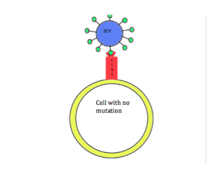
Coincidentally, research has shown that this mutation can also provide protection against HIV infection. In order for HIV to enter cells, the CCR5 receptor must be present on the surface of the cell. However, in individuals with the CCR5-∆32 mutation, the CCR5 receptor is not present on the cell membrane. Due to this mutation, individuals are resistant to HIV. This relationship between HIV and the CCR5-∆32 mutation is a way to debunk the misconception that all mutations are detrimental. In this case, having the mutation leads to increased fitness.

The protective effects of the CCR5 delta32 mutation against HIV infection are also being investigated as a potential long-term treatment option. Currently, antiretroviral therapy (ART) is the most widely used treatment for HIV, consisting of a combination of drugs that suppress progression of the disease and for reduce rates of transmission. However, ART is not a cure and individuals must rely on daily drug regimens for the entirety of their lives. Furthermore, the virus can eventually develop resistance to ART, leading to a need for more long-term treatment options.

In 2009, Hutter and other researchers identified an innovative approach using stem-cell transplantation. In a case study, a patient had HIV infection for ten years and acute myeloid leukemia. Researchers performed a stem-cell transplantation from bone marrow from an immune-compatible donor whose cells lacked expression of CCR5. The donor, who was homozygous for the CCR5 delta32 mutation, was resistant to the HIV infection. Researchers hoped that transplanting these cells with the mutation to the HIV-infected patient would confer resistance. After two transplantations, there was no recurrence of leukemia or detectable HIV in the bloodstream. The CD4+ T-cells in this patient have returned to the normal range and all carry the donor’s CCR5 gene. This patient has remained without any evidence of HIV infection for more than 8 years after discontinuation of ART, providing encouragement for stem cell transplantation as a more long-term treatment for HIV.
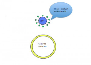
Normal cell that has the CCR5 receptor. HIV can enter and infect the cell.

Figure 2A: Normal cell that has the CCR5 receptor. HIV can enter and infect the cell.

Figure 2B: Mutated cell without the CCR5 receptor. HIV is not able to enter and infect the cell, thus making the individual immune to the virus.

Figure 2B: Mutated cell without the CCR5 receptor. HIV is not able to enter and infect the cell, thus making the individual immune to the virus.

To learn more:

Petz, L.D., et al. 2015. Progress toward curing HIV infection with hematopoietic cell transplantation. Stem Cells Cloning 8: 109-116.

Cohn, S.K., & Weaver, L.T. 2006. The Black Death and AIDS: CCR5-Δ32 in genetics and history. Q J Med 99: 497-503.

Allers, K., et al. 2011. Evidence for the cure of HIV infection by CCR5Δ32/Δ32 stem cell transplantation. Blood 117(10): 2791-99.

Galvani, A.P., & Slatkin, M. 2003. Evaluating plague and smallpox as historical selective pressures for the CCR5-Δ32 HIV-resistance allele. PNAS 100(25): 15276-79.

Shariff, Mohammed. “10 Crazy Cures for the Black Death- Listverse.” List verse. N.p., 21 Jan. 2013. Web. 06 Dec. 2015.

Libert, F., et al. 1998. The CCR5 mutation conferring protection against HIV-1 in Caucasian populations has a single and recent origin in Northeastern Europe. Human Molecular Genetics 7(3): 399-406.

Stumpf, M.P., & Wilkinson-Herbots, H.M. 2004. Allelic histories: positive selection on a HIV-resistance allele. Trends in Ecology and Evolution 19(4): 166-8.

Hutter, G., et al. 2009. Long-Term Control of HIV by CCR5 Delta32/Delta32 Stem-Cell Transplantation. New England Journal of Medicine 360: 692-698.

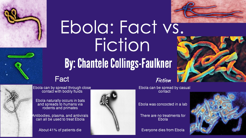
Ebola: Fact vs. Fiction

Contributed by Chantele Collings-Faulkner

We’re all going to die!!!! Right? Yes, but not necessarily from Ebola!

ebola-fact-vs-fiction-copy_20151209013526_1449624926572_block_0

Much of what is thought to be true about Ebola is actually wrong.

The genus Ebolavirus consists of five different variants of a single stranded RNA virus that originate from many parts of Africa. Though previous outbreaks have occurred in the Democratic Republic of the Congo (Olabode 2015), the most recent outbreak of Zaire ebolavirus began in Guinea and spread to Sierra Leone and Mali (Park et al 2015 and Carroll 2015). Though a series of zoonotic introductions to the human population was initially suspected to be the cause of the rapid spread of the disease, Park and colleagues determined that human to human transmission was the greatest cause of the expansion of the epidemic into Sierra Leona as the Ebola viruses shared common ancestors with a particular strain from Guinea (Park et al 2015 and Caroll 2015).

At the time, some feared the spread of Ebola to America because of the depth of interconnection between countries. However, Ebola does not spread that easily in the initial, well stages. It requires near death-bed symptoms, where caregivers are involved with caring for the sick patient. These close relatives and healthcare workers are at the greatest risk for contracting the disease, as it is spread through bodily fluids, not simple, casual contact. Symptoms of severe disease include bloody diarrhea, bloody vomiting, and bleeding through eyes, nose, mouth, and anus. Very effectively, the virus uses such fluid means to spread from person to person during care or burial, infecting others in close proximity to the patient.

Though there is no current vaccine (several are being developed), treatments include rehydration, monoclonal antibody infusion, plasma donation from survivors, and antiviral therapy called ZMapp, which has shown to be very effective in treating the disease. The Ebola virus also has not been changing genetically in the past 40 years, which offers hope for a cure (Baize 2015, Kugleman 2015, and Liu 2015). Prevention involves bleach solutions and strict adherence to PPE (personal protective equipment) guidelines to minimize exposure and reduce disease transmission.

To learn more:

Azarian, Taj, et al. 2015. Impact of spatial dispersion, evolution, and selection on Ebola Zaire Virus epidemic waves. Scientific Reports 5: 10170.

Baize, S. (2010). Towards broad protection against ebolaviruses. Future Microbiology 5: 1469-73.

Caroll, Miles W., et al. 2015. Temporal and spatial analysis of the 2014–2015 Ebola virus outbreak in West Africa. Nature 524: 97–101.

Kiran, Narasinha Mahale, Milind S. Patole. 2015. The crux and crust of ebolavirus: Analysis of genome sequences and glycoprotein gene. Biochemical and Biophysical Research Communications 463: 756–761.

Kugelman, Jeffrey R., et al. 2015. Monitoring of Ebola Virus Makona Evolution through Establishment of Advanced Genomic Capability in Liberia. Emerging Infectious Disease 21: 7.

Liu, Si-Qing, et al. 2015. Identifying the pattern of molecular evolution for Zaire ebolavirus in the 2014 outbreak in West Africa. Infection, Genetics and Evolution 32: 51–59.

Olabode, Abayomi S., et. al. 2015. Ebolavirus is evolving but not changing: No evidence for functional change in EBOV from 1976 to the 2014 outbreak. Virology 482: 202–207.

Park, Daniel J., et al. 2015. Ebola Virus Epidemiology, Transmission, and Evolution during Seven Months in Sierra Leone. Cell 161: 1516–1526.

Cancer: Evolutionary Fact or Fiction?

Contributed by Nicholas Eyrich, Jordan Feltes, Eric Ni, Somnath Das, Noah Steigelfest, and Evan Dackowski.

Cancer Heredity

Misconception: It is commonly perceived that cancer is hereditary, and one can either not have cancer in their family, thinking they are fine or have family members diagnosed and think it is a matter of time before they contract the disease.

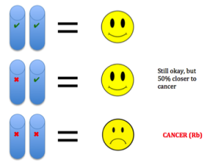
Truth: Both of these notions grossly overlook the fact that most cancers are sporadic, meaning their onset is not due to family history, but rather due to gen1etic mutations and environmental exposures during one’s lifetime. Cancer is not heritable, but a predisposition to the disease can be (Peltomaki, 2012). For example, in our DNA we have two copies of each tumor suppressor gene, one from each parent. These genes keep cells from growing uncontrollably. So to lose function, one has to have mutations in both copies. Unfortunately, one can be born with a mutation in one copy, such as in Retinoblastoma (Rb). This means the gene is still expressed using the normal copy, but this confers a 50% increased predisposition for the disease bringing cells halfway closer to being cancer cells (Price et al., 2014).

Evolutionary Cancer Mutations

Misconception: It takes one bad mutation in one cell to get cancer, and cancer is one disease with all of cells in a tumor being equal.

Truth: Population biology is used to describe tumor growth and spread (metastasis), rather than arising from a single “bad” cell. Actually, it takes on average six to seven cumulative mutations (less in pediatric cancers) to confer disease, each one having been selected for during the previous cell generation. When mutations happen and build upon each other, the combination of changes can lead to cancer. (Yamamoto, Nakamura, & Haeno, 2015). Illustrated below. This supports cancer mostly being a disease of old age, as mutations take time to accumulate. Cancer essentially consists of many diseases that continue to harass the brains of researchers. Essentially, doctors have been able to treat for certain mutations, but once one important mutation is treated, another one can take over to drive relapse (Landau et al., 2015). Also, in tumors there ar2e different environments around cells fostering different mutations in various areas of the same tumor (Hardiman et al., 2015). Cancer is adapting to therapies that target mutations, making it so difficult to control.
DNA Damage

Misconception: DNA damage is rare, and we have little protection against it. Most cancer-causing agents (carcinogens) are processed chemicals not naturally found in nature.

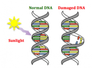
Truth: DNA damage happens many times per day and our bodies have also evolved repair mechanisms in response to the need to correct such damage. In addition, the vast majority of cancer causin3g agents are naturally occurring substances we encounter on a daily basis (Bauer, Corbett, & Doetsch, 2015). The human body has evolved ways to counteract DNA-damaging events, for example during sunlight exposure, using molecular machinery and likewise other naturally occurring compounds (Nishisgori, 2015).
 

References

Bauer, Nicholas C., Anita H. Corbett, and Paul W. Doetsch. “The Current State of Eukaryotic DNA Base Damage and Repair.” Nucleic Acids Res Nucleic Acids Research (2015): n. pag. Web.

Hardiman, Karin M., Peter J. Ulintz, Rork D. Kuick, Daniel H. Hovelson, Christopher M. Gates, Ashwini Bhasi, Ana Rodrigues Grant, Jianhua Liu, Andi K. Cani, Joel K. Greenson, Scott A. Tomlins, and Eric R. Fearon. “Intra-tumor Genetic Heterogeneity in Rectal Cancer.” Lab Invest Laboratory Investigation (2015): n. pag. Web.

Landau, Dan A., Eugen Tausch, Amaro N. Taylor-Weiner, Chip Stewart, Johannes G. Reiter, Jasmin Bahlo, Sandra Kluth, Ivana Bozic, Mike Lawrence, Sebastian Böttcher, Scott L. Carter, Kristian Cibulskis, Daniel Mertens, Carrie L. Sougnez, Mara Rosenberg, Julian M. Hess, Jennifer Edelmann, Sabrina Kless, Michael Kneba, Matthias Ritgen, Anna Fink, Kirsten Fischer, Stacey Gabriel, Eric S. Lander, Martin A. Nowak, Hartmut Döhner, Michael Hallek, Donna Neuberg, Gad Getz, Stephan Stilgenbauer, and Catherine J. Wu. “Mutations Driving CLL and Their Evolution in Progression and Relapse.” Nature 526.7574 (2015): 525-30. Web.

Nishisgori, Chikako. “Current Concept of Photocarcinogenesis.” Photochem. Photobiol. Sci. 14.9 (2015): 1713-721. Web.

Peltomäki, Päivi. “Mutations and Epimutations in the Origin of Cancer.” Experimental Cell Research 318.4 (2012): 299-310. Web.

Price, E. A., K. Price, K. Kolkiewicz, S. Hack, M. A. Reddy, J. L. Hungerford, J. E. Kingston, and Z. Onadim. “Spectrum of RB1 Mutations Identified in 403 Retinoblastoma Patients.” Journal of Medical Genetics 51.3 (2013): 208-14. Web.

Weinberg, Robert A. The Biology of Cancer. New York: Garland Science, 2007. Print.

Yamamoto, Kimiyo N., Akira Nakamura, and Hiroshi Haeno. “The Evolution of Tumor Metastasis during Clonal Expansion with Alterations in Metastasis Driver Genes.” Sci. Rep. Scientific Reports5 (2015): 15886. Web.


Helicobacter pylori and you.

Contributed by Thomas Partin and Austin Piccolo

Species do not live in a world separate from each other. Organisms interact with other organisms everyday, and over time adapt to each other accordingly. Often, the evolution of two species can become strongly linked to each other, for better or worse. Heliobacter pylori is a bacteria that thrives in the acidic conditions of the human stomach. It causes stomach ulcers and is strongly correlated with gastric cancer. As recently as the 1980’s, the idea of a bacteria being able to survive in the stomach’s harsh conditions and being responsible for this disease was so controversial that it took one doctor intentionally infecting himself to prove its role in stomach ulcers. That doctor later won the Nobel prize in medicine for his work.

H. pylori did not first start infecting humans in the 80’s though. H. pylori and humans have been living (and battling) together for millennia. The earliest humans also played host to H. pylori. One way this can be shown is a creative use of a phylogenetic tree. Scientists sampled many different strains of H. pylori, and used them to create an ancestral tree of the different strains. They then compared the tree they made to geographic locations of their samples. What they found was that lineages of H. pylori matched perfectly with the migration patterns of ancient humans as they moved out of Africa. Newer strains of H. pylori are found where humans migrated to most recently. The strains were carried and dispersed based on how early humans moved around the globe.

This intimate relation between H. pylori and humans provides a great opportunity to explore coevolution. Humans and H. pylori have been locked in an arms race for thousands of years. H. pylori colonization poses serious health consequences to the host, which creates a selective pressure for humans that can prevent H. pylori infection. Likewise, the human body is an incredibly hostile environment towards foreign invaders like H. pylori, which creates a strong selective environment for H. pylori cells that can overcome human defenses. There is evidence this selective pressure is so strong that H. pylori begins adapting specifically to the host after initial colonization. Although not an innate aspect of human biology, antibiotics are another human defense against H. pylori. Antibiotic use creates a selective pressure for H. pylori that is so strong that resistant strains can develop remarkably quickly after attempted treatment.

Please watch the below video to learn more!

https://www.youtube.com/watch?v=01NY55VV0Rg;feature=youtu.be

For further information see:

Linz, B., Ballouxm, F., Moodley, Y., Manica, A., Liu, H., Roumagnac, P., Falush, D., Stamer, C., Prugnolle, F., van der Mer, S.W., Yamaoka, Y., Graham, D.Y., Perez-Trallero, E., Wadstrom, T., Suerbaum, S., Achtman, M. 2007. An African origin for the intimate association between humans and Helicobacter pylori. Nature 445: 915-918

Gao, W., Cheng, H., Hu, F., Li, J., Wang, L., Yang, G., Xu, L., Zheng, X. 2010. The Evolution of Helicobacter pylori Antibiotics Resistance Over 10 Years in Beijing, China. Helicobacter. 15: 460-466.

Oh, J.D., Kling-Bäckhed, H., Giannakis, M., Xu, J., Fulton, R.S., Fulton, L.A., Cordum, H.S., Wang, C., Elliott, Glendoria., Edwards, J., Mardis, E.R., Engstrand, L.G., Gordon, J.I. 2006. The complete genome sequence of a chronic atrophic gastritis Helicobacter pylori strain: Evolution during disease progression. PNAS. 103: 9999-10004.

Blecker, U., Landers, S., Keppens, E., Vandenplas, Y. 1994. Evolution of Helicobacter pylori Positivity in Infants Born From Positive Mothers. Journal of Pediatric Gastroenterology and Nutrition. 19: 87-90

Kennemann, L., Didelot, X., Aebischer, T., Khun, S., Drescher, B., Droge, M., Reinhardt, R., Correa, P., Meyer, T.F., Josenhan, C., Falush, D., Suerbaum S. 2011. Helicobacter pylori genome evolution during human infection. PNAS. 108: 5033-5038.

Marshall, B.J., Warren, J.R. 1984. Unidentified curved bacilli in the stomach of patients with gastritis and peptic ulcers. The Lancet. 323: 1311-131.

Avasthi, T.S., Devi, S.H., Taylor, T.D., Kumar, N., Baddam, R., Kondo, S., Suzuki, Y., Lamouliatte, H., Mégraud, F., Ahmed, N. 2011. Genomes of Two Chronological Isolates (Helicobacter pylori 2017 and 2018) of the West African Helicobacter pylori Strain 908 Obtained from a Single Patient. Journal of Bacteriology. 193: 3385-3386.

Why You Should Thank Your Food Allergies

Contributed by Jimmy Shah, Sanjana Rao, and Laura Galarza

FOOD ALLERGY 101

Did you know that over 15 million Americans suffer from food allergies today? Consider this. Every 3 minutes, a food allergy reaction sends someone to the ER. Given these severe, potentially life-threatening medical conditions that have no known cure, we think studying the origins of food allergies can have significant clinical implications. So what exactly is a food allergy?

Food allergies manifest as adverse immune system reactions to harmless food substances. Once the allergen enters the body, it will be recognized by and bind to serum immunoglobulin E (IgE) – antibodies found in the lungs, skin, and mucosal membranes. IgE is attached by FcɛRI surface receptors on mast cells, an immune cell that helps the body create inflammatory responses. Interestingly, studies have shown that the high-affinity IgE-FcɛRI receptor binding is involved in responding to not only allergen exposure, but also parasite invasion.

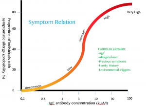
Symptom severity is correlated with IgE antibody concentration

Symptom severity is correlated with IgE antibody concentration

After the binding occurs, the allergen will cause antibody cross-linking on the mast cell surface and lead to something called mast cell degranulation, which means the mast cell will release its internal pro-inflammatory molecules, like histamines, leukotrienes, and prostaglandins, into the bloodstream. This will cause the onset of the allergy symptoms many of us may know well (especially in the pollen-rich spring) – sneezing, itching, coughing, hives, GI discomfort, etc. The most severe of these is anaphylaxis, a rapid and potentially life-threatening body state where blood pressure is lowered and emergency symptoms arise as a result. This brings us to our central question: why would evolution naturally select for us to be allergic to food that sustains us? The anomaly of food allergies and their past, present, and future benefits remain largely poorly-defined. However, research shows us that IgE antibody recognition of the allergen and shared defense mechanisms play a significant role in evolving allergic responses over time.

EVOLUTION IN FOOD ALLERGY

Although not fully understood, allergic responses are thought to have evolved from an immune defense mechanism against parasite invasion and other harmful toxin colonization. For years, scientists saw allergies as genetic accidents where aberrant IgE antibody production was just a mishap. But given the conservative nature of evolution, the IgE antibody class couldn’t have just arisen to be destructive only in the case of genetic disorders. Even if they did, evolution wouldn’t keep them around if they were solely harmful. In 1991, Margie Profet created the toxin hypothesis – the idea that responding to toxins and allergic reactions occur in very similar ways, that allergies are inherently toxic or affiliated with toxic substances, and that allergic responses mostly involve symptoms by which toxins are expelled (sneezing, vomiting, coughing, etc.) Last month, scientists at Stanford published evidence supporting the sustained positive evolutionary pressure to keep these IgE antibodies around. First, they found that in normal mice, previous exposure to venom allowed for greater survivability following a lethal venom injection, as compared to mice who were only treated with control solution. Then, they tested the role of the allergic pathway. Specifically, they studied three different types of mice responding to bee venom injections – mice without IgE, mice without IgE receptors on mast cells, and mice without mast cells at all. Unlike the normal mice, the three mutants did not benefit from previous venom exposure, since they did not have the key immunological players coordinating allergic response (Tsai, 2015). Although allergies have become less threatening in our daily lives, this allergic-type, IgE-associated immune response provides support for the idea that allergic responses are closely linked to the ways in which our bodies fight off toxins, long ago and today.

In another study published in October 2015, researchers at the London School of Hygiene & Tropical Medicine hypothesized that there must be some molecular similarity between parasites and allergen proteins, as the same branch of the immune system is found to kick in in both circumstances. After extensive data analysis, the team found that 2,445 known parasite proteins were structurally and sequentially (think base pairs A, C, T, G) similar to those found in the portion of the allergen that is prone to immune system attack. Further, measuring human cell immune response to a protein from a parasitic worm that was similar to a protein from a prevalent pollen allergen family revealed that blood serum reacted against both worm infection and the allergen via the same antibody mechanism (Tyagi, 2015).

SIGNIFICANCE

Overall, the precise origin of food allergies has yet to be defined. However, we can infer that thousands of years ago, our ancestors may have consumed foods that contained harmful proteins or mimicked harmful substances, so allergies may have very well evolved to protect us. For example, someone could have ingested a raw plant that looked like a poisonous plant in the same family, so his body was on high alert. The IgE response naturally kicked in and perhaps was sustained throughout several generations as a heritable characteristic because it gave certain individuals an advantage over others in surviving and reproducing. As a result, those advantaged individuals likely passed on these beneficial traits, so eventually the proportion of individuals with these advantageous characteristics increased because those who didn’t, would lesser survivability.

This beneficial defense mechanism is not a novel idea – if unwanted substances enter the body, whether it just appears to be harmful or actually is, the organism’s ability to survive is potentially at stake. Thus, evolution would select for mechanisms by which these substances can be fought internally and expelled from the body. It therefore makes perfect sense that evolution would select for allergic responses as a means to protect one against destructive parasites and toxins.

Ultimately, food allergies account for $25 billion dollars in health costs each year, and cause 30,000 cases of anaphylaxis, 2,000 hospitalizations, and approximately 150 deaths annually. This significant burden on our population’s health warrants study of how allergic responses occur and the reasoning behind why they do. In doing so, perhaps we can find a cure! But for now, keep in mind that food allergies aren’t necessarily all bad and that they might actually be shielding you from something far worse.

FOR FURTHER READING…

Brandtzaeg, Per. 2010. Food Allergy: Separating the Science from the Mythology. Nature Reviews Gastroenterology & Hepatology 7, no. 7: 380–400.

Fitzsimmons, Colin Matthew, Franco Harald Fakone, and David William Dunne. 2014. Helminth Allergens, Parasite-Specific IgE, and Its Protective Role in Human Immunity. Frontiers in Immunology 5: 61.

Gross, Michael. 2015. Why did evolution give us allergies? Current Biology, no. 2: 53-55.

Liu, Andrew H. 2015. Revisiting the Hygiene Hypothesis for Allergy and Asthma. Journal of Allergy and Clinical ImmMatricardi, P. M. 2014. Molecular Evolution of the Allergy. Allergologie 37, no. 10: 423–24.

Machado, D. C., Horton, D., Harrop, R., Peachell, P. T. and Helm, B. A. (1996), Potential allergens stimulate the release of mediators of the allergic response from cells of mast cell lineage in the absence of sensitization with antigen-specific IgE. Eur. J. Immunol., 26: 2972–2980. doi: 10.1002/eji.1830261224

Platts-Mills, Thomas A. E. 2012. Allergy in Evolution. New Trends in Allergy and Atopic Eczema, edited by J. Ring, U. Darsow, and H. Behrendt, 96:1–6.

Ratnaparkhe, Milind B., Tae-Ho Lee, Xu Tan, Xiyin Wang, Jingping Li, Changsoo Kim, Lisa K. Rainville, et al. 2014. Comparative and Evolutionary Analysis of Major Peanut Allergen Gene Families. Genome Biology and Evolution 6, no. 9: 2468–88.

Sicherer, Scott H., and Hugh A. Sampson. 2009. Food Allergy: Recent Advances in Pathophysiology and Treatment. Annual Review of Medicine 60, no. 1 (2009): 261–77.

Tsai, Mindy, Phillip Starkl, Thomas Marichal. 2015. Testing the “toxin hypothesis of allergy: mast cells, IgE, and innate and acquired immune responses to venoms. Elsevier. Vol. 36: 80-87.

Tyagi, Nidhi, Edward Franell, Colin Fitzsimmons, Stephanie Ryan. Comparisons of Allergic and Metazoan Parasite Proteins: Allergy of the Price of Immunity. PLOS Computational Biology.

Wang, Jing, Litao Yang, Xiaoxiang Zhao, Jing Li, and Dabing Zhang. 2014. Characterization and Phylogenetic Analysis of Allergenic Tryp_alpha_amyl Protein Family in Plants Journal of Agricultural and Food Chemistry 62, no. 1: 270–78.

Zusi, Karen. 2015. An Evolutionary Basis for Allergies. The Scientist. http://www.the-scientist.com

Evolution in Influenza

Contributed by Runako Aranha-Minnis

Evolution is not only limited to the organisms we are able to see with the naked eye. Viruses like influenza are able to evolve, and this can be very dangerous to human populations. Though it is true that influenza is not the danger that it once was, it still can not be ignored. Viral resistance to treatment can lead to many deaths globally, especially during flu seasons. During these times, doctors usually see a rise in cases of viruses resistant to the treatments available at the time. One type of resistance that influenza can develop is Oseltamivir resistance. Oseltamivir is an important antiviral defense against the flu. It is found in drugs like Tamiflu, and can be rendered ineffective if resistance is developed.

Influenza and its neuraminidase

The DNA change that in the virus that occurs is related to the neuraminidase enzyme activity. The enzyme cleaves salicylic acid moieties that can be bound by the viral hemagglutinin. In essence the enzyme’s job is to help release newly formed viral particles. Imagine the enzyme as little Pac-Men just going about and eating the connections of new virus particles to let them spread. Oseltamivir works by inhibiting the activity of the neuraminidase enzyme. This limits the infectiousness of the virus.

Resistance requires changes in the DNA of the virus. Mutations, changes in the DNA, can change genes, often leading to changes in the organism. For Oseltamivir resistance, a point mutation, the change of a single base of DNA, is all that is necessary. A change in the genes leads to a change in the amino acids produced. This change in the amino acids is just one amino acid at the 274th spot in the chain leads to a change in the activity of the virus. This line of reasoning is why the mutation is called H274Y.

The H274Y mutation that gives influenza resistance usually does not spread through the population.  In early clinical tests, the mutation meant the virus was unaffected by oseltamivir, but had associated decreased viral fitness. So even though viruses with resistance would be selected for, their lower chance of survivability because of the mutation meant it did not matter. The influenza virus is able to evolve with Oseltamivir only when an associated mutation occurs. These associated mutations that allow oseltamivir resistance to keep going are permissive mutations. More recently, permissive mutations have arisen that allow influenza to be resistant to Oseltamivir without losing any fitness. Imagine a population of influenza like a game of “Where is Waldo?”:

No permissive mutations. In the absence of permissive mutations, all viruses, including Waldo, die. 

With permissive mutations. If Waldo has a permissive mutation, he survives to reproduce and the whole populations becomes Waldos because all other viruses died. 

Waldo in the first cartoon has H274Y with no permissive mutations. Without permissive mutations, there is no evolution under treatment. After treatment, the viruses are killed. There are no viruses left so the picture goes black. In the second cartoon, With permissive mutations, there is evolution for Waldo and a new viral population arises. This population only has Waldo because only Waldo has the H274Y mutation that conferred resistance.

For Further Reading:

Jesse D. Bloom, Lizhi Ian Gong, David Baltimore. 2010. Permissive Secondary Mutations Enable the Evolution of Influenza Oseltamivir Resistance Science. 328: 1272-1275

Nicholas Renzette, Daniel R. Caffrey, Konstantin B. Zeldovich, Ping Liu, Glen R. Gallagher, Daniel Aiello, Alyssa J. Porter, Evelyn A. Kurt-Jones, Daniel N. Bolon, Yu-Ping Poh, Jeffrey D. Jensen, Celia A. Schiffer, Timothy F. Kowalik, Robert W. Finberg, Jennifer P. Wang. 2013. Evolution of the Influenza A Virus Genome during Development of Oseltamivir Resistance In Vitro 88: 272-281

The Sad History of HIV and its Persistence against Drug Therapy

Contributed by Oliver Ting, Jackson Fritz and Milan Patel

According to a report from the World Health Organization, human immunodeficiency virus (HIV) infected about 2.7 million new people in 2010 alone. Broken down, HIV destroys the immune system in humans, allowing opportunistic diseases to come in and kill the patient. HIV spreads through bodily fluid from person to person. Once inside a patient, HIV targets certain cells within the immune system called T-cells. HIV forces these cells to create new copies of the virus and destroys the T-cells in the process. As the virus grows exponentially, the immune system loses its ability to fight other diseases, leading to acquired immune deficiency virus (AIDS).

HIV cannot be cured. The main problem in creating a drug that treats HIV is that the virus is constantly changing. HIV is a retrovirus, meaning it uses RNA and then converts that into DNA when it infects cells. Retroviruses use an enzyme called reverse transcriptase to do this. However, unlike DNA enzymes, this enzyme cannot proofread itself, allowing more mutations (changes in the genetic code) to occur. This causes the virus to produce a base mismatch roughly every 10, 000–30, 000 bases during the replication. This differs significantly from regular viruses that typically have a range of one base mismatch per one million to one billion base pairs. When these base mutations occur in specific regions of the HIV DNA, new types or subtypes of the virus can be created. HIV builds up drug resistance because a drug that was effective for a previous variation of HIV may not be effective against a new variant of the virus. In addition, the virus builds cross-resistance, and becomes resistant to multiple types of HIV drugs within the same class. This further limits the number of drugs that can be used to treat the virus.

HIV is a prime example of evolution in action. HIV’s high rate of DNA mutation creates many variants. When HIV is selected against through antiviral drugs, certain variants survive that are resistant to the drug. These drug-resistant strains of HIV survive to reproduce and can be transmitted to other individuals. This constantly changing HIV population produces a substantial obstacle in trying to treat and eradicate the virus. Continued research is required to combat this evolving virus and improve the quality of life of those affected by HIV and AIDS.

Inspired by Radiolab “Patient Zero”, a fascinating tale about how HIV began, where it came from, and who “Patient 0” may have been. The podcast further reinforces how the virus has been able to combat so many challenges to its existence as well as it has.

Further Reading:

Bao, Yi, et al. “Characteristics Of HIV-1 Natural Drug Resistance-Associated Mutations In Former Paid Blood Donors In Henan Province, China.” Plos ONE 9.2 (2014): 1-9.

Bennett, Diane E . et al. “Drug Resistance Mutations for Surveillance of Transmitted HIV-1 Drug-Resistance: 2009 Update.” Ed. Douglas F. Nixon. PLoS ONE 4.3 (2009): E4724.

Sanjuan, R. et al. “Viral Mutation Rates.” Journal of Virology 84.19 (2010): 9733-748.

Shilts, Randy. And the Band Played On: Politics, People, and the AIDS Epidemic. New York: St. Martin’s, 1987.

Superbugs: The Evolution of Gonorrhea

Contributed by Nyasia Jones, Chris Richardson and Kari Tyler

https://www.youtube.com/watch?v=AesE046gtQs

A common misconception regarding evolution it that it is slow, and because it is slow, humans are not influencing it. This is completely false. Humans have and are continuing to make major changes that are not only influencing the course of our own evolution, but are also influencing the evolution of other species we interact with. Especially in medicine, human advancement is occurring at an amazing pace and thus allowing us to witness evolution in response to our actions.

Over the past century, the use of antibiotics to treat bacterial pathogens has become a widespread practice. Starting around 1930, medical practitioners began discovering several drugs that successfully rid patients of a particular bacterial pathogen Neisseria gonorrhoeae. The first successful therapy was administration of a class of chemicals called sulfonamides. For years, treatment with sulfonamides proved successful in patient after patient until about the mid-1940s when reports of N. gonorrhoeae resistance to sulfonamides increased. Fortunately, at this time, the “miracle drug” penicillin was found to be highly effective at bacterial treatment and become the number one treatment option. After ten to fifteen years though, low doses of penicillin were no longer as effective, and by the 1980s strains with high-level penicillin resistance had emerged.

So what happened with both penicillin and the sulfonamides?  Nothing.

But something did happen to the N. gonorrhoeae. They evolved.

Sequential chromosomal mutations allowed some bacteria to incur resistance to penicillin. With penicillin treatment, the bacteria with resistance survived while those without it did not. The resistant individuals reproduced thereby creating a new generation of bacteria in which all individuals were penicillin resistant. This, my friends, is evolution.

Unlike the billions and billions of years it took to create modern-day humans, this evolution took less than a century to change these N. gonorrhoeae bacteria into “superbugs” which are becoming increasingly harder to treat. And it doesn’t stop there. N. gonorrhoeae has also become resistant to more recent treatment options, such as tetracycline and fluoroquinolones. Now, with less and less success to current methods of treatment, namely cefixime and ceftriaxone, scientists are worried history will repeat itself and strains of N. gonnorhoeae with complete cefixime and ceftriaxone resistance will emerge. With dwindling options for treatment, N. gonorrhoeae resistance and that of other superbugs remains a major problem in the fields of medicine and epidemiology.

So are humans influencing evolution? N. gonorrhoeae tell us, loud and clear: yes!

For more information:

Anderson, M. T., & Seifert, H. S. (2011). Neisseria gonorrhoeae and humans perform an evolutionary LINE dance. Mobile Genetic Elements, 1(1), 85-87.

Mavroidi, A., Tzelepi, E., Siatravani, E., Godoy, D., Miriagou, V., & Spratt, B. G. (2011). Analysis of emergence of Quinolone-resistant gonococci in Greece by combined use of Neisseria gonorrhoeae multiantigen sequence typing and multilocus sequence typing. Journal of Clinical Microbiology, 49(4), 1196-1201.

Unemo, M., & Shafer, W. M. (2011). Antibiotic resistance in Neisseria gonorrhoeae: Origin, evolution, and lessons learned for the future. Annals of the New York Academy of Sciences, 1230(1), E19-E28.

Venom Variation

Contributed by Annelise Bonvillian, Cherisma Patel and Liz Pinkerton

I introduce to you two rattlesnakes. I think they’re generally friendly but they could also be planning to maim me. You just never know with venomous snakes. 

Judging by their appearance, you’d think both rattlesnakes were of the same species. You’d be right, being the clever person you are. They are in fact part of the same subspecies of Southern Pacific Rattlesnake (Crotalus oreganus helleri).

These C. o. helleri rattlesnakes are venomous. Before we dive into their story, let’s explain venom.

Venom Across The Tree of Life

Many animals have evolved venom for protection or to help them capture prey. We, unfortunately have not.

There are also a wide range of structures for delivering venom, such as the cute little fangs in these rattlesnakes.

The interesting thing is, similar types of venom have evolved in completely unrelated species. This is not because all venomous species evolved from one super venomous ancestor or because the venomous critters of the world get together to talk strategy.

Evolution of venom is a solid example of convergent evolution in which organisms that are not very closely related independently evolve similar traits due to similar environmental pressures. The proteins in the toxin end up acting on the same physiological molecule by chance. Most venoms involve disrupting major physiological pathways that are accessible by the bloodstream, especially the hemostatic and neurological systems. Damaging these systems would efficiently cripple any prey or potential predator.

Back to the Rattlesnakes

Getting back to our original C. o. helleri rattlesnakes, you’d think it might be true that they’d have the same type of venom because they’re of the same subspecies, right?

In fact, these two rattlesnakes are very closely related, but the populations that live in the mountains have a distinctly different venom than those that live in the desert.

Distribution of Crotalus oreganus

Distribution of Crotalus oreganus

A research team from the University of Queensland studied the variation between rattlesnakes in the mountainous area of Idyllwild, California and those in the desert of Phelan, California. The extent of venom variation is surprising for two populations because the two areas are relatively close together geographically.

The venom of the desert-dwelling rattlesnake contains proteins that break down blood vessels and prevent clotting.

This venom is slow acting. It is described as haemotoxic and would lead to uncontrollable bleeding. Neat!

Its close relative in the mountains is equally as morbid. The mountain populations have proteins in their venom that stop nerves from sending signals to the muscles.

This venom is fast acting. It is described as neurotoxic and results in paralysis. Sweet!

The venoms are completely different even though these two populations are from the same subspecies.

In fact, venom can vary between species, subspecies, individuals, sexes, and ages. Another research team from the Biomedical Institute of Valencia that loves poisonous reptiles just as much as the the previous one studied venom variability in Mojave rattlesnakes in Arizona. They found that you were 10 times more likely to die from a bite in one county than another. Here, the severity, not the type, of venom was what varied.

So, getting back to our C. o. helleri rattlesnakes (don’t worry, we love you best), here’s another question: which would be better to have in the snake’s arsenal? The fast acting neurotoxin venom or the slow acting haemotoxin?

Evolution, the lovely process that developed these fine creatures is NOT about having the best of a trait. The fitness of a certain trait is relative. What is good for one population might not necessarily be the best for another. Based on where they live, different venom types might be more beneficial in capturing prey or warding off predators.

So why would it be more beneficial for the mountain rattlesnake to have a faster acting venom?

Researchers suspect that environmental differences between these two populations of rattlesnake are likely to have promoted the huge variation in venom between the two.

If the mountain rattlesnake’s venom was slow acting the, prey could hide before the venom had properly incapacitated it.

And venom isn’t cheap. Creating venom costs a lot of energy and wasting it time and time again would be a shame. Natural selection would thus lead to a venom composition that would reduce metabolic cost. Natural selection is the process by which organisms that have higher fitness and are more adapted to their environment tend to produce more offspring and survive.

Now, in the desert, rattlesnakes don’t have to worry as much about their prey hiding before they can get to them. They might not have evolved the quicker neurotoxic venom because there was no selective pressure to have their venom be fast acting.

As with most things in this incomprehensible universe, we are not sure if the desert dwelling Southern Pacific rattlesnake maintains its haemotoxic venom due to lack of selection pressure. Or perhaps somehow the snakes have co-evolved with their prey and the haemotoxic venom is just the most efficient one for the prey type.

All in all, it’s important to realize that evolution isn’t a race where the end goal is to have the best of something. Traits change randomly due to mutations, and factors such as the environment or prey type select for variations that are most beneficial for survival.

In related news, using the vast powers of human intelligence to learn from the natural world, snake venom is being adapted to heal instead of hurt. Venom works in the same way as many medicines, and the enzymes in venom are being modified to affect disease processes.

One specific example includes the using ACE inhibitors in Brazilian pit viper venom to prevent hypertension.

However, with the decline of snake diversity due to environmental degradation, the diversity of venom and its medical potential is decreasing.The fact that venom from various snakes can be used to target certain  diseases is a very important implication for evolutionary medicine. Population divergence in snakes increases the potential for variation in venom type, which can ultimately increase the antidotes possible for fighting certain diseases.

So why research things like rattlesnake venom? Not only is the subject wildly fascinating, but unraveling the complexities of snake venom can help humans better counter its life-threatening effects and can also promote the development of new medicines. In conclusion,  though we may be terrified of you, dear rattlesnakes and other venomous denizens of this world, we’d also like to say thanks. May you continue to amaze us.

Check out these sites for more information:

Caswell, Nocholas R., Wolfgang Wuster, Freek J. Vink, Robert A. Harrison, and Bryan G. Fry. 2012. Complex cocktails: the evolutionary novelty of venoms Trends in Ecology and Evolution.

Holland, Jennifer S. 2013. Venom: The Bite That Heals. National Geographic: The New Age of Exploration. http://ngm.nationalgeographic.com/2013/02/125-venom/holland-text

Kartik Sunagara, Eivind A.B. Undheimc, Holger Scheibd, Eric C.K. Grene, Chip Cochrane, Carl E. Persone, Ivan Koludarovc, Wayne Kellne, William K. Hayese, Glenn F. Kingd, Agosthino Antunesa, Bryan Grieg Fry. Intraspecific venom variation in the medically significant Southern Pacific Rattlesnake (Crotalus oreganus helleri): Biodiscovery, clinical and evolutionary implications. 2014. Journal of Proteomics. http://www.sciencedirect.com/science/article/pii/S1874391914000256

Massey DJ, Calvete JJ, Sánchez EE, Sanz L, Richards K, Curtis R, Boesen K. 2012. Venom variability and envenoming severity outcomes of the Crotalus scutulatus scutulatus (Mojave rattlesnake) from Southern Arizona. Journal of Proteomics. http://www.ncbi.nlm.nih.gov/pubmed/22446891

Yong, Ed. 2014. Rattlesnakes Two Hours Apart Pack Totally Different Venoms. National Geographic: Phenomena. Online. http://phenomena.nationalgeographic.com/2014/01/27/rattlesnakes-two-hours-apart-pack-totally-different-venoms/

Zimmer, Carl. 2013. On the Origin of Venom. National Geographic: Phenomena. Online. http://phenomena.nationalgeographic.com/2013/01/09/on-the-origin-of-venom/

Rise of Antibiotic Resistance

Contributed by Richard Parilla

https://www.youtube.com/watch?v=gXisYehVXUk

In 1928 a discovery made by Scottish scientist Alexander Fleming would change modern medicine. Fleming observed that the fungus Penicillium was able to kill disease causing pathogens. This discovery won Fleming a Nobel Prize in 1945 and led to development of antibiotics. The effectiveness of these drugs, once referred to as “miracle drugs”, led them to become used very regularly to treat a variety of illnesses. However, these lifesaving antibiotics have become far less effective due to the increasingly more common bacterial strains that are antibiotic resistant.

Antibiotics function by either directly killing bacteria themselves or by inhibiting their growth and reproduction. So how does bacterial strains resistance to certain antibiotics come about? Well an easy way to think about this is in evolutionary terms. The use of antibiotics selects for bacterial strains that are resistant to the mechanism by which the antibiotic targets the pathogen. Since all the non-resistant bacteria are wiped out by the antibiotic, only the resistant bacteria can reproduce and spread. This is an interesting perspective since it shows that humans can have an impact on the evolution of a species through implementation of novel selective pressures such as antibiotics.

Over the past decade antibiotic resistance has become a very threatening problem to society. It is has been a topic which has continually made headlines over the past few years. Why has the threat of antibiotic resistant microbes risen? The World Health Organization points to the misuse of antibiotics and over prescription as the cause of the accelerated emergence of the resistant bacteria strains. Due to the nature of this problem it would seem that it will be a frequent topic of discussion in the near future.

For Further Further Reading:

“Antimicrobial Resistance.” WHO. N.p., May 2013. Web. 20 Apr. 2014.

Austin, D. J. “The Relationship between the Volume of Antimicrobial Consumption in Human Communities and the Frequency of Resistance.” Proceedings of the National Academy of Sciences 96.3 (1999): 1152-156. Print.

Bonhoeffer, S. “Evaluating Treatment Protocols to Prevent Antibiotic Resistance.” Proceedings of the National Academy of Sciences 94.22 (1997): 12106-2111. Print.
Centers for Disease Control and Prevention. Centers for Disease Control and Prevention, 16 Sept. 2013. Web. 20 Apr. 2014.

“General Background: About Antibiotic Resistance.” Tufts University. N.p., n.d. Web. 20 Apr. 2014.

Hampton, L. M., M. M. Farley, W. Schaffner, A. Thomas, A. Reingold, L. H. Harrison, R. Lynfield, N. M. Bennett, S. Petit, K. Gershman, J. Baumbach, B. Beall, J. Jorgensen, A. Glennen, E. R. Zell, and M. Moore. “Prevention of Antibiotic-Nonsusceptible Streptococcus Pneumoniae With Conjugate Vaccines.”Journal of Infectious Diseases 205.3 (2012): 401-11. Print.

Levy, Stuart B. “The Challenge of Antibiotic Resistance.” Diss. Texas U, 1998. Web.

Lupo, Agnese, Sébastien Coyne, and Thomas Ulrich Berendonk. “Origin and Evolution of Antibiotic Resistance: The Common Mechanisms of Emergence and Spread in Water Bodies.” Frontiers in Microbiology 3 (2012): n. pag. Print.

Stöppler, Melissa C., M.D. “Antibiotics 101 – MedicineNet.com.” MedicineNet. N.p., 3 Sept. 2012. Web. 20 Apr. 2014.

Woodford, Neil, Jane F. Turton, and David M. Livermore. “Multiresistant Gram-negative Bacteria: The Role of High-risk Clones in the Dissemination of Antibiotic Resistance.” FEMS Microbiology Reviews 35.5 (2011): 736-55. Print.